Close is not close enough
Scientists unravel important mechanism for fast communication between neurons
Avoiding hitting a running child on the street or reacting rapidly to an alarm call – timely reactions are only possible because our neurons communicate extremely fast with each other. Scientists from Braunschweig and Göttingen have now discovered a decisive mechanism that allows for such fast signal transmissions.

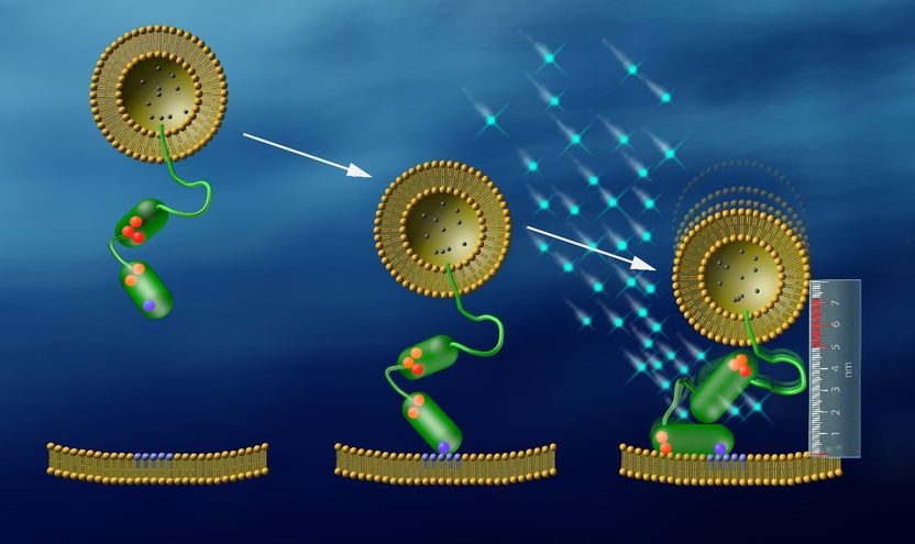
Using fluorescence signals and a nanoruler consisting of DNA, Peter J. Walla and his co-workers were able to observe exactly how synaptotagmin (green) tethers vesicles to membranes. With a distance of eight nanometers the vesicles are just ready to start the communication between neurons. As soon as the calcium level increases, calcium ions (light blue) bind to synaptotagmin, which then pulls the vesicle to a distance of five nanometers from the membrane. At this distance vesicles can fuse with the membrane and release the neurotransmitters.
© Walla, Max Planck Institute for Biophysical Chemistry
Communication is not only of vital importance for social life – cells are also continuously interacting with each other, thus allowing us to breathe, move, and think. Without rapid signal transmission between neurons, even the world class keeper Manuel Neuer would not be able to make a single save. Usually, the signals are carried by neurotransmitter molecules. In small membrane packages – so-called synaptic vesicles – they await at the ends of a neuron a calcium signal to take action. As soon as the signal arrives, the message is delivered by fusion of many synaptic vesicles with the cell membrane of the sending neuron, thereby releasing the neurotransmitters. Sequentially, these neurotransmitters activate a corresponding signal in the receiving neuron. It was well known that this process is started by an increase of calcium concentration in the sending neuron. However, the triggering process of how the vesicles actually recognize and process this signal just at the right time was very poorly understood.
Fusion of vesicles and membranes of the sending cells is only possible when they are in close proximity. Peter J. Walla and a team of researchers at the University of Braunschweig and the Max Planck Institute (MPI) for Biophysical Chemistry in Göttingen were now able to observe a protein decisive for this process directly “at work“. “This protein – called synaptotagmin – is able to tether the vesicles with the membrane at exactly the proper distances. Synaptotagmin can be regarded as a kind of molecular anchor”, explains Walla, Professor at the University of Braunschweig and head of the Research Group Biomolecular Spectroscopy and Single Molecule Detection at the MPI for Biophysical Chemistry.
Synaptotagmin is anchored to the vesicles at one end. The scientists now observed that it tethers with the other end to the membrane at a distance of about eight nanometers (one millionth of a millimeter). In this stage the vesicles are ready to take action very quickly when required. “But the distance is still large enough to prevent fusion of vesicles with the membrane and the resulting release of the neurotransmitters“, Walla states. This is because the so-called SNARE proteins on both vesicle and membrane, which govern the fusion process itself, need to catch each other like in a zipper. However, at a distance of eight nanometers this zipper is simply unable to close completely.
Once the calcium concentration increases, this gives the starting signal. “First, the calcium binds to synaptotagmin. Immediately after that, the protein pulls the vesicle and membrane to a distance of only five nanometers. This is a distance at which the SNAREs can quickly fuse the membranes“, the scientist explains. “Our work is the first evidence ever showing that synaptotagmin can change its conformation upon a calcium trigger in a way that decreases the distance between vesicles and membranes just on the proper scales.“
To date, there simply had been no appropriate tools to measure the exact distance between vesicles and membranes. The research team around Walla has now succeeded by developing a new, elaborate technique: They used DNA-strands of very well defined lengths as a ruler on the nanometer scale. By observing changing light intensities and durations from fluorescence markers, the scientists could determine the distance between vesicles and the membrane with an accuracy of around one nanometer. “Measuring short distances between two single points using fluorescence markers is a common approach. However, measuring distances also between surfaces of entire membranes with the required precision was only possible since we introduced the concept of DNA-based nanorulers”, says Chao-Chen Lin at the MPI for Biophysical Chemistry.
The new nanoruler for membrane distances might proof a very useful tool also in important fields other than neurobiology. Peter J. Walla is convinced: “For example, this tool can also be used in biological processes like the infection of cells by viruses or in the fertilization of eggs. In these cases distances between membranes also play a vital role”.


