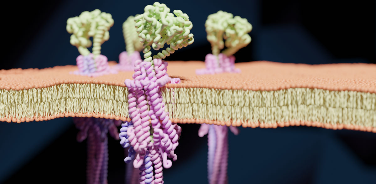
A stronghold in the fight against viruses - new bacterial immune system decoded
International research team describes for the first time the structure and function of the Zorya system
bacteria are constantly infected by viruses, so-called phages, which use the bacteria as host cells. However, in the course of evolution, bacteria have developed various strategies to protect themselves from these attacks. Many of these bacterial immunity systems have been known for a long time. Prof. Dr Marc Erhardt and Prof. Dr Philipp Popp, both from the Institute of Biology at Humboldt-Universität zu Berlin, together with colleagues in Denmark and New Zealand and other partners, have now unveiled the structure and function of a novel bacterial defence system against phages. It was discovered in 2018 by an Israeli research group and named after Zorya, a figure in Slavic mythology. The research results have now been published in the scientific journal Nature.
The Zorya system recognises phage attacks and activates an early and precise defence that destroys the phage DNA while avoiding the death of the host cell. “Zorya is like an early warning system with a protective shield. It recognises the first signs of an attack and reacts very fast to fend off the intruder,” explains Prof Marc Erhardt, head of the Molecular Microbiology Lab at Humboldt-Universität zu Berlin and one of the lead authors of the study.
Stronghold against phages
The investigation of the Zorya system using state-of-the-art methods such as cryo-electron and fluorescence microscopy shows that it consists of an intricate molecular motor and several specialised components. This motor senses at an early stage changes in the cell wall caused by penetrating phages and triggers a series of protective reactions. This previously unknown mechanism enables the bacterial cell to degrade the phage DNA so that the virus cannot multiply in the host cell. This is remarkable because bacteria usually prevent phages from multiplying by inducing cell death or – in other words – ‘sacrificing’ themselves. “Decoding the Zorya system was like opening a treasure chest,” says Erhardt. “You keep discovering new facets of this molecular masterpiece.”
To analyse the structure of the protein complexes, samples were cooled down to very low temperatures of up to -260 °C within fractions of a second using cryo-electron microscopy. This shock freezing prevents the formation of ice crystals so that molecules keep their natural form. Fluorescence microscopy, in turn, provided an insight into the interaction of the virus particles with the bacterial cells.
New possibilities for biotechnological applications
The decoding of this antiviral system of bacteria has far-reaching implications: On the one hand, it contributes to a better understanding of the mechanisms of phage-bacteria interaction. On the other hand, the findings open up new avenues for biotechnological applications. “The Zorya system could serve as a basis for the development of innovative tools for the precise manipulation of genetic material or for the development of novel therapies against bacterial infections,” adds Prof Philipp Popp, visiting professor at the Department of Biology and co-author of the study. The development of the Nobel Prize-winning CRISPR-Cas method for genome editing is also based on a bacterial immunity system that protects against viruses, discovered in the 2000s. For Philipp Popp, the current study is also an example of the beauty of molecular biology: “It is fascinating to see how elegant the survival strategies developed by bacteria are. Zorya shows us how much we can still learn about these tiny but incredibly complex organisms.”
Original publication
Haidai Hu, Philipp F. Popp, Thomas C. D. Hughes, Aritz Roa-Eguiara, Nicole R. Rutbeek, Freddie J. O. Martin, Ivo Alexander Hendriks, Leighton J. Payne, Yumeng Yan, Dorentina Humolli, Victor Klein-Sousa, Inga Songailiene, Yong Wang, Michael Lund Nielsen, Richard M. Berry, Alexander Harms, Marc Erhardt, Simon A. Jackson, Nicholas M. I. Taylor; "Structure and mechanism of the Zorya anti-phage defense system"; Nature, 2024-12-11
See the theme worlds for related content
Topic world Fluorescence microscopy
Fluorescence microscopy has revolutionized life sciences, biotechnology and pharmaceuticals. With its ability to visualize specific molecules and structures in cells and tissues through fluorescent markers, it offers unique insights at the molecular and cellular level. With its high sensitivity and resolution, fluorescence microscopy facilitates the understanding of complex biological processes and drives innovation in therapy and diagnostics.


