Dividing walls: How immune cells enter tissue
Findings give the full picture of a process just as important for healing as for the spread of cancer
To get to the places where they are needed, immune cells not only squeeze through tiny pores. They even overcome wall-like barriers of tightly packed cells. Scientists at the Institute of Science and Technology Austria (ISTA) have now discovered that cell division is key to their success. Together with other recent studies, their findings published in Science magazine give the full picture of a process just as important for healing as for the spread of cancer.

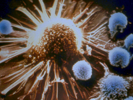
Dividing walls: How immune cells enter tissue (symbolic image).
Unsplash
Imagine a stone wall in the countryside. Tightly packed, one stone sits on top of the other filling the tiniest gaps. A seemingly unbreachable obstacle. On their way throughout the body to fight infections, immune cells face such barriers in the form of cell-dense tissues. To do their job as the body’s rescue service, they need to find a way through. In a recent study, scientists from ISTA’s Siekhaus group together with collaborators from the European Molecular Biology Laboratory (EMBL) and three students from a local High School, took a close look at how this happens in fruit fly embryos.
During the development of these tiny, transparent animals, macrophages, the dominant form of immune cells in fruit flies, infiltrate tissues. Using high-end microscopes, the scientists were able to follow their journey. “The macrophages arrive at the wall and look for the right place to enter,” explains Maria Akhmanova, until recently a postdoc at Daria Siekhaus’ research group and first author of the study.
Breaking new ground
Cues that guide the macrophages have directed them to the right spot. There, the pioneer macrophage, the first cell to move in, is waiting. Suddenly, a part of the wall starts to move. The cell right in front of the macrophage rounds up, preparing to divide – a normal part of its cell cycle. “This is what the pioneer has been waiting for,” says Akhmanova. Moving its cell nucleus ahead, the pioneer cell now pushes forward while all the other macrophages follow in its tracks. As the Siekhaus group also recently discovered, to break through the pioneer gets an extra boost of energy through a complex process governed by a newly discovered protein the scientists named Atossa. Furthermore, the scientists learned that to shield their sensitive nucleus from damage, the macrophages develop protective armor made from actin filaments.
Cell division crucial for success
By precisely inhibiting, slowing down, and speeding up the division specifically of the flanking tissue cells, the researchers were now able to prove that the crucial component that allows immune cells to enter is in fact surrounding cell division. As it rounds up to prepare for division, the tissue cell at the entry site loses some of its connection points to its surroundings, the researchers observed through live imaging. In collaboration with the De Renzis lab at EMBL, the researchers also artificially induced rounding through a cutting edge technique using light to induce genetic changes. This wasn’t sufficient to get the macrophages to enter. But genetically reducing the amount of the cell connections was. “It was very exciting to see how the macrophages were only able to enter the tissue when the tissue cell lost its connections,” says Akhmanova.
Powerful implications for cancer research
“Cell division being the key process that controls macrophage infiltration is really a very elegant concept with powerful implications,” Professor Daria Siekhaus enthuses. The same mechanism that helps macrophages enter tissues could also be essential for many other types of immune cells in vertebrates like humans. In the long run, the scientists are eager to learn if manipulating the connections or the divisions of the tissue cells could help increase immune cells’ infiltration of tumors to fight them from within or help reduce immune cells’ ability to attack tissues during autoimmunity. “Our findings will also affect any researcher who is working on any migrating cell in the context of the body,” the cell biologist explains.
For her study, the theoretical biophysicist and Lise Meitner fellow Maria Akhmanova delved deep into the world of microscopy. With the help of her mentor Daria Siekhaus, she learned everything she could about the fascinating and very helpful fruit flies. Three students from the Klosterneuburg High School were also part of the team. During a school trip to the Institute's laboratories, they discovered their enthusiasm for research. Consequently they helped Akhmanova with crossing and identifying fruit flies and even wrote an algorithm to speed up image analysis. “The success of this research project was made possible by joint forces from many scientists and enormous help from three motivated high school students!” says Akhmanova.


