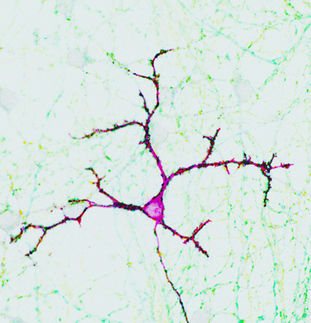
The hunt for mirror neurons – not every technique picks them up
Researchers in Tübingen have been studying how mirror neurons, which are assumed to be key to the understanding of behaviour, respond when the same action is repeatedly observed. They found answers in the cerebral cortex of monkeys. The study has a surprising result: the mirror neuron system does not adapt. This contradicts the original assumption of researchers, that mirror neurons – the same as other nerve cells – react to the frequent repetition of a particular stimulus through a reduced level of activity (adaptation.)
The results of the study highlight the importance of finding a new interpretation of neuroimaging studies that have so far shown that mirror neurons adapt. Astonishingly, these studies had shown adaptation. Researchers at the Hertie Institute for Clinical Brain Research (HIH) and the Werner Reichardt Centre for Integrative Neuroscience at the University of Tübingen have found an explanation for these apparently contradictory results.
Mirror neurons respond to goal-directed behaviours
As with other nerve cells, it is possible to stimulate mirror neurons, which can transmit this stimulation on to other nerve cells. This happens with the help of electrical impulses, which ‘fire’ up to several hundred times a second. This ‘firing’ can be measured by an electrode. Researchers had previously discovered that mirror neurons control hand movements that are directed towards a particular goal, for instance grasping a piece of apple. Yet what is special about mirror neurons is that they are similarly active when these kinds of goal-directed actions are merely observed. Hence they might play a decisive role in comprehending the behaviour of other people.
Unexpected pattern of activity baffles researchers
‘Surprisingly, it was shown that two thirds of mirror neurons did not adapt their firing patterns, as had previously been assumed’, says Pomper, a scientist at the Hertie Institute for Clinical Brain Research (HIH). Studies carried out with functional Magnetic Resonance Imaging (fMRI) had shown the opposite: the mirror neurons adapted, that is they reacted more and more weakly to the repetition of the same stimulus.
Not every approach is able to measure
The activity measured by fMRI is only able to record the ‘firing’ of nerve cells indirectly. It merely identifies changes in the blood flow through oxygen levels in the red blood cells. Experts describe this as the BOLD effect. They are evoked by the energy needs of active nerve cells. Input signals, certain processing stages in the cell protruberances (dendrites) and cell bodies, along with the activity of glial cells, another element of the nervous system, also contribute to this. ‘‘As a result, conclusions about the behavior of individual cells cannot be drawn directly from changing in BOLD signal’ says Dr. Vittorio Caggiano, formerly at Hertie Institute for Clinical Brain Research and Center for Integrative Neuroscience, University Clinic Tübingen(1).The assumption up to now has always been that an adaptation of nerve cells was the basis of the reduction of the BOLD effect seen in experiments when the same action is subsequently performed and then observed. According to the authors of the study, on the basis of the results of the current study the former interpretation of the BOLD adaptation does not stand up.
So how to explain the BOLD adaptation? One interpretation the researchers can offer is the additional raised local field potentials (LFP) in the brain: these do in fact manifest the anticipated adaptation and so they could explain the fMRI data. In every nerve cell the signal transfer goes through input signals and occasionally output signals, so-called action potentials. If the input signal to be converted into an output signal, that would lead to the firing of neurotransmitters. These in turn stimulate the next nerve cell along as a transfer signal. The researchers suggest that LFP express the input signals that come from other areas of the brain and the expression of them in a particular region.
New interpretation of the current study
It has always been that mirror neurons fire in the same way regardless of whether a subject personally performs an action or watches it. Therefore they seemed to be verifiable when one compares the fMRI signals from the execution and the observation of actions, whether of the same kind or different. ‘A one-to-one correspondence of fMRI data and the activity of mirror neurons is thus not possible. Hence from our point of view the need to find a new account of the neuro-imaging studies based on adaptation’ is ho it is summarized by Peter Their, a member of the HIH board and Chairman of the CIN.


