How skin cells protect themselves against stress
Cell biologists have developed a new method for measuring how mechanical forces in cells are processed
Advertisement
The skin is our largest organ, and, among other things, it provides protection against mechanical impacts. To ensure this protection, skin cells have to be connected to one another especially closely. Exactly how this mechanical stability is provided on the molecular level was unclear for a long time. Researchers in the team led by Prof. Carsten Grashoff from the Institute of Molecular cell biology at the University of Münster and the Max Planck Institute of Biochemistry have been collaborating with colleagues at Ludwig Maximilian University of Munich and Stanford University in the USA, and they are now able to demonstrate how mechanical stress on specialized adhesion points, so-called desmosomes, is processed. They designed a mini-measuring device, which can determine forces along individual components of the desmosomes. In the study, published in “Nature Communications”, they show how mechanical forces propagate along these structures.

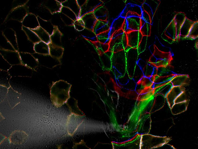
The molecular transmission of force in desmosomes was studied before (blue), during (green) and after (red) application of mechanical stress.
WWU - AG Grashoff
Cells in the skin stick together
Our skin acts as a protective shield against external influences and has to withstand very different stresses. It has to be able to stretch but must not tear when exposed to great strains. To fulfil this mechanical function, skin cells form specialized adhesion points, so-called desmosomes, which strengthen the adhesion between cells. Patients with deficient desmosomes suffer from severe skin disorders, which arise after the exposure to mechanical stress. What was hitherto barely understood, however, was how mechanical forces impact on the individual components of the desmosomes. The international group of researchers has developed a method for analysing the molecular forces at these adhesion points.
Miniature spring balance measures force in desmosomes
“This technique functions in way that is similar to a miniature spring scale,” says Anna-Lena Cost from the Max Planck Institute, who is one of the lead authors of the study. The force sensor consists of two fluorescent dyes, which are connected with an extensible peptide. The peptide acts as a spring, which is stretched by just a few piconewtons – which in turn leads to a change in the dyes’ radiance. The researchers are able to read this change with a microscope so that mechanical differences at individual binding points can be determined. In their experiments, the researchers discovered that desmosomes are not exposed to any mechanical stress as long as external forces are absent. If cells are pulled – as it frequently happens in the skin – then mechanical stress becomes apparent in the desmosomes. These forms of stress depend on the force magnitude and its orientation. “When there is only a low level of mechanical stress, other structures in the cell can carry the burden. But if a high degree of stress occurs, then desmosomes come to the rescue,” summarizes Anna-Lena Cost.


