Researchers isolate mouse heart stem cell
Isolation, tracking of mouse stem cells ends debate on their existence
A pioneering Cornell and University of Bonn study has isolated and purified mouse heart stem cells, settling a debate over whether such cells exist. The findings, published in Proceedings of the National Academy of Sciences, could allow researchers to better understand whether genes can spur heart stem cells to fully differentiate into new cells after a heart attack.
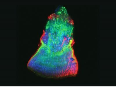
The researchers, led by Michael I. Kotlikoff, the Austin O. Hooey Dean of Veterinary Medicine at Cornell, used a green fluorescent protein to label mouse heart precursor (or stem) cells and identify the cells during embryo development and immediately following birth. The fluorescent protein label also revealed that the number of cells, which differentiate into all three heart cell types (cardiac, endothelial and smooth muscle), decline drastically soon after birth.
The new method could be used to quickly and rapidly isolate and purify both heart and other stem cell populations in the laboratory; to study gene expression that leads to these cells differentiating into other cell types; to track the timing of these cells and when and where they differentiate into other cell types in vivo; and to compare heart stem cells with other types of stem cells.
"The existence of cardiac stem cells and the ability of adult stem cells to form new heart muscle have been the subject of much scientific disagreement, as there are so few of these cells in the adult heart," said Kotlikoff, who co-authored the study with Yvonne Tallini, a Cornell research scientist in biomedical sciences, and Bernd Fleischmann of the University of Bonn.
"We now have a simple way to identify these cells within the heart and to isolate and study the factors that control their fate," Kotlikoff added.
Researchers had questioned the existence of these cells, because the heart has very little regenerative capacity after an infarction, which creates a permanent scar. To address this question, the group looked for the cells after heart infarction, and found that heart stem cells form vessels that invade the scar tissue, but do not form new heart cells. Heart cells surrounding the dead tissue express a protein marker for these stem cells at low levels, suggesting that they are attempting to respond to grow new heart cells after an injury, but the response is incomplete. This may explain the detection of these cells after an injury, but their failure to re-grow new heart tissue.
Other news from the department science


Most read news


More news from our other portals


Last viewed contents
Journal_of_Bone_&_Joint_Surgery
Category:Marine_biologists

Ionic and covalent drug delivery
Bothriopsis
Evotec Completes Divesture of Chemical Development