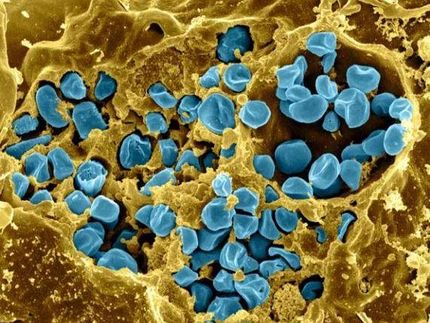
Cornell researchers study bacterium big enough to see
Advertisement
The secret to an unusual bacterium's massive size - it's the size of a grain of salt, or a million times bigger than E. coli bacteria, and big enough to see with the naked eye - may be found in its ability to copy its genome tens of thousands of times. That's according to Cornell research published in Proceedings of the National Academy of Sciences.
This giant among bacteria, Epulopiscium sp., lives in a symbiotic relationship in the gut of surgeonfish around Australia's Great Barrier Reef. The research shows how a simple modification in the basic design of bacterial cells allows Epulopiscium sp. to grow so large.
"Other bacteria have multiple copies of their genome, but prior to this, I think the highest numbers known have been a hundred or a few hundred copies," said Esther Angert, a Cornell associate professor of microbiology and the paper's senior author. "The big discovery is seeing this bacterium with tens of thousands of copies of its genome."
Most bacteria are small and appear to be structurally simple. They lack the specialized organelles that allow eukaryotic cells (cells in which DNA is contained within a nucleus) to take in nutrients, organize cellular functions and maintain larger sizes. Bacteria instead rely on diffusion through their cell membranes to obtain nutrients and other important chemicals. Since bacteria cannot move nutrients within the cell body, they need to stay small for diffusion to work well.
But, by copying its genome thousands of times and arraying it in a kind of fabric just under the cell membrane, Epulopiscium sp. may maintain its large size by keeping its DNA close to the outer surface, Angert said. That way, the DNA may respond quickly and locally to stimuli by producing RNA and proteins where they are needed.
"Having copies of its genome arrayed around the periphery keeps the DNA close to the outer environment," said Angert. "The bacterium can immediately react as something comes in contact with the cell."
The bacterium's large size offers advantages: It is highly mobile and too big to eat for most protozoan predators that also live in the surgeonfish's gut.
Also, while most bacteria reproduce by dividing into two equal-sized offspring, Epulopiscium sp. produces offspring internally, usually two, one at each pole of the cigar-shaped cell. These polar cells grow within the mother cell's cytoplasm, until the mother cell eventually bursts open and dies.
"We're interested in how that process arose and how that may affect the biology of the organism," said Angert.


