Visible signals from brain and heart
New sensor measures calcium concentrations deep inside tissue
Key processes in the body are controlled by the concentration of calcium in and around cells. A team from the Technical University of Munich (TUM) and the Helmholtz Zentrum München have developed the first sensor molecule that is able to visualize calcium in living animals with the help of a radiation-free imaging technique known as optoacoustics. The method does not require the cells to be genetically modified and involves no radiation exposure.

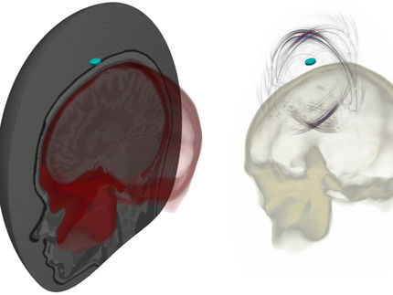
Calcium waves – a new sensor converts light to sound to visualize calcium fluxes in the body.
B. van Rossum / G. Westmeyer
Calcium is an important messenger in the body. In nerve cells, for example, calcium ions determine whether signals are relayed to other nerve cells. And whether a muscle contracts or relaxes depends on the concentration of calcium in the muscle cells. This is also true of the most vital muscle in our body – the heart.
“Because calcium plays such an important role in essential organs such as the heart and brain, it would be interesting to be able to observe how calcium concentrations change deep within living tissues and in this way to improve our understanding of disease processes. Our sensor molecule is a small first step in this direction,” says Gil Gregor Westmeyer, head of the study and Professor of Molecular Imaging at TUM and Research Group Leader at Helmholtz Zentrum München. In the study, which was published in in the Journal of the American Chemical Society, Professor Thorsten Bach of the TUM’s Department of Chemistry was also involved. The researchers have already tested their molecule in the heart tissue and brains of living zebra fish larvae.
Calcium measurements also possible in deep tissue
The sensor can be measured using a relatively new, non-invasive imaging method known as optoacoustics, which makes it suitable for use in living animals – and later possibly also in humans. The method is based on ultrasound technology, which is harmless for humans and uses no radiation. Laser pulses heat up the photoabsorbing sensor molecule in tissue. This causes the molecule to expand briefly, resulting in the generation of ultrasound signals. The signals are then sensed by ultrasound detectors and are translated into three-dimensional images.
As light passes through tissue, it is scattered. For this reason, images under a light microscope become blurred at depths of less than a millimeter. This highlights another advantage of optoacoustics: ultrasound undergoes very little scattering, producing sharp images even at depths of several centimeters. This is particularly useful for examining the brain, because existing methods only penetrate a few millimeters below the brain surface. But the brain has such a complex three-dimensional structure with various functional areas that the surface only makes up a small part of it. The researchers therefore aim to use the new sensor to measure calcium changes deep inside living tissue. They have already achieved results in the brains of zebrafish larvae.
Nontoxic and radiation-free
Additionally, the scientists have designed the sensor molecule so that it is easily taken up by living cells. Moreover, it is harmless to tissues and works based on a color change: as soon as the sensor binds to calcium, its color changes which in turn changes the light-induced optoacoustic signal.
Many imaging methods for visualizing calcium changes that are currently available require genetically modified cells. They are programmed, for example, to fluoresce whenever the calcium concentration in the cell changes. The problem with this, of course, is that it is not possible to carry out such genetic interventions in humans.
The new sensor overcomes this limitation, the scientists say. In the future, the researchers plan to refine the properties of the molecule further, allowing the sensor signals to be measured in even deeper tissue layers. To this end, the team headed by Gil Gregor Westmeyer must generate further variants of the molecule that absorb light of a longer wavelength than cannot be perceived by the human eye.


