Researchers zero-in on cholesterol's role in cells
Scientists have long puzzled over cholesterol. It's biologically necessary; it's observably harmful - and nobody knows what it's doing where it's most abundant in cells: in the cell membrane.

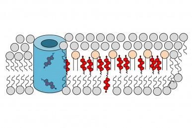
This is a diagram showing cholesterol's (red) predominance in the outer layer of the cell membrane. Cholesterol transport proteins (blue) can alter its distribution between the inner and outer layers.
UIC
Now, for the first time, chemists at the University of Illinois at Chicago have used a path-breaking optical imaging technique to pinpoint cholesterol's location and movement within the membrane. They made the surprising finding that, in addition to its many other biological roles, cholesterol is a signaling molecule that transmits messages across the cell membrane.
"Cholesterol is a lipid that gets bad press because of its association with cardiovascular disease," says Wonhwa Cho, professor of chemistry at UIC, who led the research. "It's been very well studied, but not much is known about its cellular function. What is its role? Is it a bad lipid? Absolutely not - for example, the brain is about half lipid, and cholesterol is the richest lipid in the brain," he said. A cholesterol deficiency can cause several diseases, and the substance is the starting material for making the body's dozen or so steroid hormones.
Cho's earlier studies showed cholesterol interacts with many regulatory molecules - mostly cellular proteins - but it was never thought to be one.
"We knew it could play an important role in cell regulation - for example, in proliferation or development," he said. "We know that high-fat diets, which boost cholesterol levels, have been linked to an elevated incidence of cancer. How, is not fully understood," Cho said.
One of the biggest problems conceptually, he said, is that a regulatory or signaling lipid should exist only transiently to transmit the message.
"But cholesterol is there all the time," he said. The membrane contains up to 90 percent of a cell's total cholesterol, and cholesterol makes up about 40 percent of the membrane lipids.
Cholesterol lends stability to the membrane, which is actually a double layer of lipid - or fat - molecules. The cholesterol gathers into "rafts," which were thought to serve as platforms from which other signaling molecules might operate.
"But in this paper, we showed that a single cholesterol molecule can itself be the signal trigger," Cho said.
Until now, scientists believed cholesterol was in both layers of the membrane, Cho said, "maybe more in the inner layer. But we, for the first time, measured cholesterol levels in the inner and outer layers simultaneously in real time, in living cells. And we showed that cholesterol is predominantly in the outer layer."
Cholesterol makes up about 40 percent of the outer layer of the membrane, they found, and only about 3 percent of the inner layer. In response to a specific cell stimulus, the amount in the inner layer more than doubles, and the level in the outer layer drops by the same amount.
They also found that, while in normal cells the concentration of cholesterol in the inner layer is low, in cancer cells it's much higher. "We checked this in many different cell lines," Cho said.
The new study sheds some light on the positive side effect of statin drugs lowering cancer risk. Cho and his coworkers found that treating cells with a statin dramatically lowered the level of cholesterol in the inner layer, leading to suppression of cell growth activity. This suggests a new way to treat cancer through pharmacological modulation of the cellular cholesterol level, Cho said.
"I think we're just scratching the surface of the regulatory role of cholesterol. We have many unpublished data indicating that cholesterol is involved in a wide variety of cellular processes and regulation," he said.
Lipids like cholesterol are "very nasty molecules to work with," Cho says, because they can't be dissolved in water like most biological molecules. This makes quantitative techniques very challenging.
"We had to devise a new strategy," he said. Six years ago, he and his colleagues developed an optical imaging technology that allows direct quantification of lipids in living cells. They tagged a lipid-binding protein molecule with a fluorescent sensor that changes color when it binds lipid. The color-change indicates the ratio of bound to free lipid, letting them determine how much of the lipid is at a given location in the cell membrane.
Original publication
Shu-Lin Liu, Ren Sheng, Jae Hun Jung, Li Wang, Ewa Stec, Matthew J O'Connor, Seohyoen Song, Rama Kamesh Bikkavilli, Robert A Winn, Daesung Lee, Kwanghee Baek, Kazumitsu Ueda, Irena Levitan, Kwang-Pyo Kim & Wonhwa Cho; "Orthogonal lipid sensors identify transbilayer asymmetry of plasma membrane cholesterol"; Nature Chemical Biology; 2016


