Alzheimer's researchers find clues to toxic forms of amyloid beta
A subtle change affects its aggregation behavior and stabilizes a form with enhanced toxicity
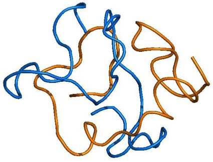
Much of the research on Alzheimer's disease has focused on the amyloid beta protein, which clumps together into sticky fibrils that form deposits in the brains of people with the disease. In recent years, attention has turned away from the fibrils themselves to an intermediate stage in the aggregation of amyloid beta. "Oligomers" consisting of a few molecules of the protein stuck together are more mobile than the large, insoluble fibrils and seem to be much more toxic. But the actual structure of these soluble oligomers remains unknown, and it's unclear how they trigger the neurotoxic effects that lead to Alzheimer's disease.
A new study by researchers at UC Santa Cruz may help lift the veil on the structure and behavior of these neurotoxic oligomers. The researchers made a subtle alteration to the amyloid beta protein that had striking effects on its properties. By replacing one amino acid with its mirror image, they created a version of amyloid beta with a reduced rate of fibril formation, different fibril structure, and increased toxicity in cell culture compared to the normal or "wild type" protein.
"We perturbed the system very slightly and got enhanced cytotoxicity and destabilization of fibrils," said Jevgenij Raskatov, assistant professor of chemistry and biochemistry at UC Santa Cruz.
Most amino acids can occur in two mirror-image forms, a "left-handed" or L-form and a "right-handed" or D-form, but living cells only make proteins out of L-amino acids. Raskatov's team changed one amino acid in the amyloid beta protein to its D-form: the glutamate at position 22.
"This subtle change stabilizes a prefibrillary intermediate that has higher toxicity," he said, noting that the stable intermediate could be a very useful tool for investigating the neurotoxic effects of amyloid beta oligomers. The role of the amyloid beta protein in Alzheimer's disease is complicated and remains poorly understood. With a better understanding of the molecular mechanisms underlying the disease, researchers hope to identify new targets for drug development efforts.
Several mutations associated with inherited early-onset Alzheimer's disease affect position 22 in the amyloid beta protein, either changing it from glutamate to another amino acid or deleting it. The new findings underscore the importance of the amino acid at position 22 for amyloid beta toxicity.
Raskatov's team--postdoctoral researchers Christopher Warner (first author) and Subrata Dutta and graduate student Alejandro Foley--used a variety of techniques to investigate the properties of the altered amyloid beta protein and the oligomers and fibrils it forms. Using small-angle x-ray scattering analysis, they found evidence that the protein forms a soluble oligomer with an ordered structure.
"The scattering experiment provided an indication of structure, and there is a chance we can use this information to gain some structural insights," Raskatov said. "We're pretty excited about that, because if we can understand the structure of the neurotoxic oligomers, that could help efforts to design molecules to disrupt them."
Original publication
Other news from the department science

Get the life science industry in your inbox
By submitting this form you agree that LUMITOS AG will send you the newsletter(s) selected above by email. Your data will not be passed on to third parties. Your data will be stored and processed in accordance with our data protection regulations. LUMITOS may contact you by email for the purpose of advertising or market and opinion surveys. You can revoke your consent at any time without giving reasons to LUMITOS AG, Ernst-Augustin-Str. 2, 12489 Berlin, Germany or by e-mail at revoke@lumitos.com with effect for the future. In addition, each email contains a link to unsubscribe from the corresponding newsletter.
Most read news
More news from our other portals
Last viewed contents
Siraitia_grosvenorii

Squeezing low-cost electricity from sustainable biomaterial
Candida albicans digs transcellular tunnels

Right under your nose: A more convenient way to diagnose Alzheimer's disease - Certain proteins in nasal discharge can indicate the onset and progression of Alzheimer's, providing an avenue for early detection

Pangolin the inspiration for medical robot - It could one day reach even the narrowest and most sensitive regions in the body in a minimally invasive and gentle way
Ciclosporin

Laser activated gold pyramids could deliver drugs, DNA into cells without harm
Category:Fellows_of_the_Royal_College_of_Physicians

Long-Sought Workaround to Helium Shortage? - Neutron Detection Method
Category:Bicyclic_antidepressants
Horses_in_warfare


