Crystal clear images uncover secrets of hormone receptors
NIH researchers gain better understanding of how neuropeptide hormones trigger chemical reactions in cells
Many hormones and neurotransmitters work by binding to receptors on a cell's exterior surface. NIH scientists used atomic level images to show how the neuropeptide hormone neurotensin might activate its receptors. Their description is the first of its kind for a neuropeptide-binding G protein-coupled receptor (GPCR).
"G protein-coupled receptors are found throughout the body. Knowing how they work should help scientists devise better treatments," said Reinhard Grisshammer, Ph.D., at the NIH's National Institute of Neurological Disorders and Stroke (NINDS)
Neurotensin is thought to be involved in Parkinson's disease, schizophrenia, temperature regulation, pain, and cancer cell growth. Previously, Dr. Grisshammer and his colleagues showed how neurotensin binds to the part of its receptor located on a cell's surface. In this study, they demonstrated how binding changes the structure of the rest of the receptor, which passes through a cell's membrane and into its interior. There neurotensin receptors activate G proteins, a group of molecules inside cells that controls a series of chemical chain reactions.
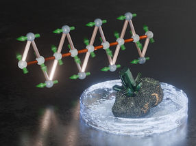
For these experiments, scientists shot X-rays at crystallized neurotensin receptor molecules. Making crystals of receptors that activate G proteins is difficult. In most studies, scientists have investigated inactive receptors.
"The receptor we crystallized is very close to the active form found in nature," said Dr. Grisshammer. "We may have the first picture of a peptide-binding G protein-coupled receptor just before it engages with the G protein."
To achieve their results, the scientists made multiple genetic modifications to a less active version of the neurotensin receptor they had used before. Experiments performed in test tubes showed that mixing the receptor with neurotensin sparked the G protein reactions for which the scientists were looking.
When the scientists looked at the structure of the new crystals, they discovered how binding of neurotensin to the receptor caused critical parts of the receptor located below a cell's surface to change shape. In particular, they saw that a region in the middle of the receptor dropped like a draw bridge to link the neurotensin binding site to parts of the receptor found inside cells that are important for G protein activation. The scientists concluded that this change may prepare the receptor for activating G proteins.
Other news from the department science


Most read news


More news from our other portals


Last viewed contents
Santhera Initiates Collaboration with the European Vision Institute Clinical Research Network in LHON

Regular caffeine consumption does not result in extra heartbeats
Evotec Extends Medicinal Chemistry Collaboration with Ono Pharmaceutical
Start of Phase IIa study in acute lung injury patients marks development milestone for Apeiron's rhACE2
Biocitech announces the arrival of BioBank for Life as new resident
PAION AG: Court sanctions Scheme of Arrangement for acquisition of CeNeS
DYNA Instruments GmbH - Hamburg, Germany
Cerebrovascular_disease
Intercell obtains results from its first Phase I investigational vaccine study for Streptococcus pneumoniae
List_of_prehistoric_cartilaginous_fish