How memories form: A glimpse into the 3D brain
Advertisement


People who wish to know how memory works are forced to take a glimpse into the brain. They can now do so without bloodshed: RUB researchers have developed a new method for creating 3D models of memory-relevant brain structures. They published their results in the trade journal “Frontiers in Neuroanatomy”.
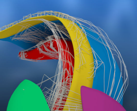
3D image of the hippocampus of a rat
© M. Pyka
Sea Horse gave the hippocampus the name
The way neurons are interconnected in the brain is very complicated. This holds especially true for the cells of the hippocampus. It is one of the oldest brain regions and its form resembles a sea horse (hippocampus in Latin). The hippocampus enables us to navigate space securely and to form personal memories. So far, the anatomic knowledge of the networks inside the hippocampus and its connection to the rest of the brain has left scientists guessing which information arrived where and when.
Signals spread through the brain
Accordingly, Dr Martin Pyka and his colleagues from the Mercator Research Group have developed a method which facilitates the reconstruction of the brain's anatomic data as a 3D model on the computer. This approach is quite unique, because it enables automatic calculation of the neural interconnection on the basis of their position inside the space and their projection directions. Biologically feasible network structures can thus be generated more easily than it used to be the case with the method available to date. Deploying 3D models, the researchers use this technique to monitor the way neural signals spread throughout the network time-wise. They have, for example, found evidence that the hippocampus’ form and size could explain why neurons in those networks fire in certain frequencies.
Information become memories
In future, this method may help us understand how animals, for example, combine various information to form memories within the hippocampus, in order to memorise food sources or dangers and to remember them in certain situations.
Original publication
Pyka M, Klatt S and Cheng S (2014): Parametric Anatomical Modeling: A method for modeling the anatomical layout of neurons and their projections, Front. Neuroanat. 8:91.


Other news from the department science


