Motional layers in the brain
Neurobiologists discover elementary motion detectors in the fruit fly
Recognising movement and its direction is one of the first and most important processing steps in any visual system. By this way, nearby predators or prey can be detected and even one’s own movements are controlled. More than fifty years ago, a mathematical model predicted how elementary motion detectors must be structured in the brain. However, which nerve cells perform this job and how they are actually connected remained a mystery. Scientists at the Max Planck Institute of Neurobiology in Martinsried have now come one crucial step closer to this “holy grail of motion vision”: They identified the cells that represent these so-called "elementary motion detectors" in the fruit fly brain. The results show that motion of an observed object is processed in two separate pathways. In each pathway, motion information is processed independently of one another and sorted according to its direction.

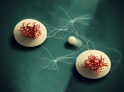
In the brains of fruit flies, specific neurons are tuned for directional information about a movement and pass this information into separate, independent layers of the brain.
© MPI of Neurobiology / Borst
Ramón y Cajal, the famous neuroanatomist, was the first to examine the brains of flies. Almost a century ago, he thus discovered a group of cells he described as “curious elements with two tufts”. About 50 years later, German physicist Werner Reichardt postulated from his behavioural experiments with flies that they possess “elementary motion detectors”, as he referred to them. These detectors compare changes in luminance between two neighbouring photoreceptor units, or facets, in the fruit fly’s eye for every point in the visual space. The direction of a local movement is then calculated from this. At least, that is what the theory predicts. Since that time, the fruit fly research community has been speculating about whether these “two-tufted cells” described by Cajal are the mysterious elementary motion detectors.
The answer to this question has been slow in coming, as the tufted cells are extremely small – much too small for sticking an electrode into them and capturing their electrical signals. Now, Alexander Borst and his group at the Max Planck- Institute of Neurobiology have succeeded in making a breakthrough with the aid of a calcium indicator. These fluorescent proteins are formed by the neurons themselves and change their fluorescence when the cells are active. It thus finally became possible for the scientists to observe and measure the activity of the tufted cells under the microscope. The results prove that these cells actually are the elementary motion detectors predicted by Werner Reichardt.
As further experiments have shown, the tufted cells can be divided into two groups. One group (T4 cells) only reacts to a transition from dark to light caused by motion, while the other group (T5 cells) reacts oppositely – only for light-to-dark edges. In every group there are four subgroups, each of which only responds to movements in a specific direction – to the right, left, upwards or downwards. The neurons in these directionally selective groups release their information into layers of subsequent nerve tissue that are completely separated from one another. There, large neurons use these signals for visual flight control, generating the appropriate commands for the flight musculature, for example. This could be impressively proven by the scientists: When they blocked the T4 cells, the neurons connected downstream and the fruit flies themselves were shown in behavioural tests to be blind to motions caused by dark-to-light edges. When the T5 cells were blocked, light-to-dark edges could no longer be perceived.
In discussions about their research results, which have just been published in the scientific journal Nature, both lead authors, Matt Maisak and Jürgen Haag, were very impressed with the “cleanly differentiated, yet highly ordered” motion information within the brains of the fruit flies. Alexander Borst, head of the study, adds: “That was real teamwork – almost all of the members in my department took part in the experiments. One group carried out the calcium measurements, another worked on the electrophysiology, and a third made the behavioural measurements. They all pulled together. It was a wonderful experience.” And it should continue like this, since the scientists are already turning to the next mammoth challenge: they would now like to identify the neurons that deliver the input signals to the elementary motion detectors. According to Reichardt, the two signals coming from neighbouring photoreceptors in the eye have to be delayed in relation to one another. “That is going to be really exciting!” says Alexander Borst.
Original publication
Matthew S Maisak, Jürgen Haag, Georg Ammer, et al., A directional tuning map of Drosophila elementary motion detectors Nature, 8 August 2013


