Polymer ribbons for better healing
Freiburg researchers develop hydrogels for tissue regeneration that can be fine-tuned to fit any body part
Advertisement
A new kind of gel that promotes the proper organization of human cells was developed by Prof. Prasad Shastri of the Institute of Macromolecular Chemistry and BIOSS Centre for Biological Signalling Studies Excellence Cluster at the University of Freiburg and BIOSS Centre for Biological Signalling Studies graduate students Aurelien Forget and Jon Christensen in collaboration with Dr. Steffen Lüdeke of the Institute for Pharmaceutical Sciences. These hydrogels made of agarose, a polymer of sugar molecules derived from sea algae, mimic many aspects of the environment of cells in the human body. They can serve as a scaffold for cells to organize in tissues. In the cover article of the Proceedings of the National Academy of Sciences Prof. Shastri and co-workers show how by applying these hydrogels they could grow blood vessel structures from cells in an unparalleled way. These gels could be used in the future to help damaged tissue heal faster.

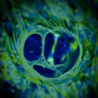
3-D organization and branching of human endothelial cells into vascular trees in carboxylated agarose gels
© Aurelien Forget, Prasad Shastri
The cells environment in the body is composed of collagen and polymers of sugars. It provides mechanical signals to the cells, necessary for their survival and proper organization into a tissue, and hence essential for healing. A gel can mimic this scaffold. However it has to precisely reproduce the molecular matrix outside the cell in its physical properties. Those properties, like the matrices stiffness, vary in the body depending on the tissue.
The team of Prof. Shastri modified agarose gels by adding a carboxylic acid residue to the molecular structure of the polymer to optimally fit the cells environment. Hydrogels form when polymer chains that can dissolve in water are crosslinked. In an agarose gel the sugar chains organize into a spring-like structure. By adding a carboxylic acid to this backbone, the polymers form ribbon-like structures – this allows for the stiffness of the gel to be tuned to adapt the scaffold to every part of the human body.
To demonstrate the versatility of the gel the researchers manipulated endothelial cells that make up vascular tissue to organize into blood vessels outside the body. By combining the appropriate biological molecules found in a developing embryo, they identified a single condition that encourages endothelial cells to form large blood vessel-like structures, several hundred micrometers in height. This discovery has implications in treating damage to heart and muscle tissue.
Prof. Shastri says “it is really remarkable that the organization of the endothelial cells into these free standing vascular lumens occurs within our gels without the need for support cells”. It has been long thought the formation of large vessel-like structures requires additional cells called mural support cells, which provide a platform for the endothelial cells to attach and organize. “We were surprised to find that the endothelial cells underwent a specific transformation called apical-basal polarization”, adds Prof. Shastri. It turns out that such polarization is necessary for the development of blood vessels and occurs naturally in a developing embryo. The ability to induce this polarization in cells in three-dimensional cultures in a synthetic polymer environment is a unique feature of the new gel.


