Dental X-rays linked to common brain tumor
Research finds correlation between frequent dental X-rays and increased risk of developing meningioma
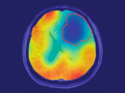
Meningioma, the most common primary brain tumor in the United States, accounts for about 33 percent of all primary brain tumors. The most consistently identified environmental risk factor for meningioma is exposure to ionizing radiation. In the largest study of its kind, researchers from Brigham and Women's Hospital (BWH), Yale University School of Medicine, Duke University, UCSF and Baylor College of Medicine have found a correlation between past frequent dental x-rays, which are the most common source of exposure to ionizing radiation in the U.S, and an increased risk of developing meningioma. These findings are published in Cancer.
"The findings suggest that dental x-rays obtained in the past at increased frequently and at a young age, may be associated with increased risk of developing this common type of brain tumor," said Elizabeth Claus, MD, PhD, a neurosurgeon at BWH and Yale University School of Medicine at New Haven. "This research suggests that although dental x-rays are an important tool in maintaining good oral health, efforts to moderate exposure to this form of imaging may be of benefit to some patients."
Claus and her colleagues studied data from 1,433 patients diagnosed with meningioma between 20 and 79 years of age between May 2006 and April 2011 and compared the information to a control group of 1350 participants with similar characteristics. They found that patients with meningioma were twice as likely to report having a specific type of dental x-ray called a bitewing exam, and that those who reported having them yearly or more frequently were 1.4 to 1.9 times as likely to develop a meningioma when compared to the control group. Additionally, researchers report that there was an even greater increased risk of meningioma in patients who reported having a panorex x-ray exam. Those who reported having this exam taken under the age of 10, were 4.9 times more likely to develop a meningioma compared to controls. Those who reported having the exam yearly or more frequently than once a year were nearly 3 times as likely to develop meningioma when compared to the control group.
"It is important to note that the dental x-rays performed today use a much lower dose of radiation than in the past," said Claus.
According to background information in the study, The American Dental Association's statement on the use of dental radiographs emphasizes the need for dentists to examine the risks and benefits of dental x-rays and confirms that there is little evidence to support the use of dental x-rays in healthy patients at preset intervals.
Most read news
Other news from the department science

Get the life science industry in your inbox
By submitting this form you agree that LUMITOS AG will send you the newsletter(s) selected above by email. Your data will not be passed on to third parties. Your data will be stored and processed in accordance with our data protection regulations. LUMITOS may contact you by email for the purpose of advertising or market and opinion surveys. You can revoke your consent at any time without giving reasons to LUMITOS AG, Ernst-Augustin-Str. 2, 12489 Berlin, Germany or by e-mail at revoke@lumitos.com with effect for the future. In addition, each email contains a link to unsubscribe from the corresponding newsletter.
Most read news
More news from our other portals
Last viewed contents
Waldemar_Haffkine
Solomon_H._Snyder

How cells recognize uninvited guests
IKBKAP

New separation process for key radiodiagnostic agent reduces radioactive waste - Less waste from lower enriched Uranium targets
Mace_(spray)
Cardiopulmonary_bypass


