Stretched, ordered DNA molecules could bring insights into disease
Studying chemical modifications in the chromosomes of cells is akin to searching for changes in coiled spaghetti. Scientists at Cornell have figured out how to stretch out tangled strands of DNA from chromosomes, line them up and tag them to reflect different levels of modification -- which could lead to insights into how these chemical processes affect human health.
Researchers in the lab of Harold Craighead, the Charles W. Lake Professor of Engineering, used advanced nanofabrication techniques to make it easier to see how single molecules of DNA subtly change during a chemical process called methylation. A better understanding of this process could lead to further study into genetic machinery of numerous diseases, including Alzheimer's, Parkinson's, diabetes and cancer.
This work was published online Oct. 7 in the journal Analytical Chemistry.
DNA is normally packed tightly into the nucleus of a cell and housed into chromosomes. Within chromosomes lie many chemical perturbations that modify how genes are expressed, without anything to do with the underlying DNA sequence, explained Aline Cerf, a postdoctoral associate who led the study.
Studying these chemical modifications is a relatively new field called epigenetics. The researchers focused on a particular process in which a section of the DNA, called 5-cytosine, becomes methylated -- its chemical structure changes by the addition of a methyl group (CH3).
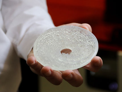
Cerf and colleagues first took genetic material extracted from cells and suspended it in solution. Using a technique called soft lithography, they made a stamp, consisting of micrometer-sized wells, out of polydimethylsiloxane (PDMS). The solution of coiled DNA molecules was deposited onto the PDMS stamp, which was then moved at a controlled speed across a glass surface. The molecules became trapped by capillary forces in the wells, and stretched.
The result was an ordered array of elongated molecules that were transferred by contact onto a support to be imaged and studied.
In related work, the researchers also published a paper in the journal Nano Letters, Sept. 16, which details how their molecular arrays could be transferred to and imaged on substrates of graphene -- single-layer sheets of carbon atoms. This let them get even better pictures of the molecules by allowing the use of transmission electron microscopy for imaging of the added tags and the underlying base sequence in the DNA.
"The DNA molecule arrays can be tagged, separated and lined up to quickly read off the genetic and epigenetic information," Craighead said.
Researchers have in the past studied these same molecules while still contained in their nuclei, he said, but this new technique simplifies that process.
"We're no longer dealing with bundles of molecules that are hindering the information of another one," Craighead said. "We are taking the equivalent of the DNA contained in one cell and spreading it out on a surface, so you can look at all of these things and search for and study things of interest that will no longer be lost in this disordered world we were in before. Aline's results have brought us out of that world."
The studies were supported by the National Cancer Institute and the National Institutes of Health.
Most read news
Topics
Organizations
Other news from the department science

Get the life science industry in your inbox
By submitting this form you agree that LUMITOS AG will send you the newsletter(s) selected above by email. Your data will not be passed on to third parties. Your data will be stored and processed in accordance with our data protection regulations. LUMITOS may contact you by email for the purpose of advertising or market and opinion surveys. You can revoke your consent at any time without giving reasons to LUMITOS AG, Ernst-Augustin-Str. 2, 12489 Berlin, Germany or by e-mail at revoke@lumitos.com with effect for the future. In addition, each email contains a link to unsubscribe from the corresponding newsletter.
Most read news
More news from our other portals
Last viewed contents
Study uncovers new hurdle for developing immunotherapies
How a molecular Superman protects the genome from damage - Scientists find a new role for RNAi protein Dicer in preventing collisions during DNA replication
Networks of Gene Activity Control Organ Development
AIDS_advocacy

Evotec completes acquisition of Rigenerand
