The Nervous System as a Three-Dimensional Map
Freiburg Research Team Creates First Complete Map of Special Connections of Nerve Cells in Zebrafish
It would be hard to understand how a city is organized without knowledge of the course of all of its streets. Scientists confront the same problem when trying to understand the functioning of the brain. In the case of vertebrates, we only have fragmentary knowledge of which nerve cells send their connections, so-called axons, into certain regions of the brain. Such knowledge is particularly important for understanding the functioning of groups of nerves that send out axons to modulate the activity of neural circuits in remote regions of the brain. One of these groups consists of nerve cells that use the tiny molecule dopamine as a messenger to control many types of behavior. It is precisely these neurons which die off in those afflicted with Parkinson’s disease, a fact which demonstrates the central role they play in medicine.

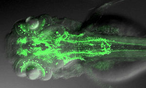
Coloured overall view of a Zebrafish nervous system
Driever and Ryu
A team of neurobiologists from the University of Freiburg, led by Prof. Dr. Wolfgang Driever from the Faculty of Biology and including Dr. Olaf Ronneberger from the Department of Computer Science and Dr. Roland Nitschke from the Center for Systems Biology (ZBSA), has now succeeded in creating the first complete map of all axons which use dopamine as a messenger in a vertebrate, namely in the model organism zebrafish. The data identify all projection possibilities, the so-called “projectome,” of every nerve cell for a class of messengers in the nervous system which is of great importance for medicine. The researchers cooperated closely with the university’s Center for Systems Biology (ZBSA) and BIOSS, Centre for Biological Signalling Studies.
The scientists were able to create the first three-dimensional projectome map of the intact brain of the zebrafish by combining the selective genetic marking of individual nerve cells with high resolution microscopy at the ZBSA. The new map reveals important information on the possible functioning of the brain. For instance, it illustrates that dopaminergic neurons of the diencephalon connect distant regions of the brain in previously unimagined ways – regions responsible for higher brain functions in the telencephalon, physiological control in the hypothalamus, the coordination of movement in the hindbrain and the execution of movement in the spinal cord. These neurons can be involved in effecting changes in basic behavioral states following stress: active reactions like fight or flight or passive reactions like freezing all activity. In the same study, the scientists describe a new dopaminergic system in another region of the zebrafish’s brain, the corpus striatum, in which the loss of dopaminergic connections in Parkinson patients is particularly severe. The authors speculate that this system might compensate for the low amount of dopaminergic neurons in fish. In conjunction with further neurobiological studies, the “projectom” map opens up possibilities for a new understanding of neural circuits in the brains of simple vertebrates like the zebrafish.
Original publication
Most read news
Original publication
Tuan Leng Tay, Olaf Ronneberger, Soojin Ryu, Roland Nitschke, and Wolfgang Driever. (2011) Comprehensive catecholaminergic projectome analysis reveals single neuron integration of zebrafish ascending and descending dopaminergic systems. Nature Communications 25 January 2011
Organizations
Other news from the department science

Get the life science industry in your inbox
By submitting this form you agree that LUMITOS AG will send you the newsletter(s) selected above by email. Your data will not be passed on to third parties. Your data will be stored and processed in accordance with our data protection regulations. LUMITOS may contact you by email for the purpose of advertising or market and opinion surveys. You can revoke your consent at any time without giving reasons to LUMITOS AG, Ernst-Augustin-Str. 2, 12489 Berlin, Germany or by e-mail at revoke@lumitos.com with effect for the future. In addition, each email contains a link to unsubscribe from the corresponding newsletter.


