Same types of cell respond differently to stimulus, Stanford study shows
Using new technology that allows scientists to monitor how individual cells react in the complex system of cell signaling, Stanford University researchers have uncovered a much larger spectrum of differences between each cell than ever seen before. Cells don't all act in a uniform fashion, as was previously thought.
"Think of cells as musicians in a jazz band," said Markus Covert, PhD, assistant professor of bioengineering and senior author of the study, which will be published online in Nature June 27. Covert's lab studies complex genetic systems. "One little trumpet starts to play, and the cells go off on their own riffs. One plays off of the other."
Up to now, most of the scientific information gathered on cell signaling has been obtained from populations of cells using bulk assays due to technological limitations on the ability to examine each individual cell. The new study, using an imaging system developed at Stanford based on microfluidics, shows that scientists have been misled by the results of the cell-population-based studies.
"While the outcome of activation may be the same, the process the cells use to achieve this outcome is very different," the study authors wrote. "Population studies have not revealed the intricate network of information one observes at the single cell level."
"This really surprised us," said study co-author Stephen Quake, PhD, a professor of bioengineering at Stanford, investigator of the Howard Hughes Medical Institute and a leader in the field of microfluidics. "It sends us back to the drawing board to figure out what is really going on in cells."
Cell signaling governs basic cellular activities and coordinates cell actions in the human body. The ability of cells to correctly respond to their environments is the basis of all development, tissue repair and immunity. A better understanding of how cells talk to each other could lead to new insights into how larger biological systems operate, and possibly lead to cures for such diseases as cancer, diabetes and autoimmune disorders, which are caused by errors in this process.
"What we see is that differences between cells matter," Covert said. "Even the nuances can play a role."
To achieve his goal of studying individual cell reactions during the cell-signaling process, Covert's lab joined forces with Quake's lab.
Quake, who is also the Lee Otterson Professor in the School of Engineering, had invented the biological equivalent of the integrated circuit — the microfluidic chip — which enables a single researcher to achieve what once would have required dozens or more. Three years ago, researchers in his lab, Rafael Gomez-Sjoberg and Annel Leyrat, developed a microfluidic chip specifically for the study of single cells. In this study, Quake and Covert put it to use to investigate inflammatory cell signaling.
"This study is a beautiful biological application of microfluidic cell culture and really illustrates the power of the technology," Quake said.
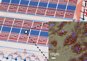
The chip is made of three layers of a silicon-based clear elastic material and contains the microscopic equivalent of test tubes, pipettes and petri dishes. Valves and gates control fluid flow. By regulating flow, the chip carries out dozens of experiments at the same time. It's essentially a lab on a chip.
"We used a microfluidics platform that could maintain and monitor cell cultures 96 at a time," Covert said. "I was doing one at a time before that. Over a one-year period, we were able to study, with unprecedented detail, how 5,000 cells responded to signals. This took us to a totally new dimension."
The scientists put mouse fibroblast cells onto the chip and let them grow in an environmental chamber, which is mounted on an inverted microscope. The entire system, which fits on a small desktop, is computerized and provides long-term monitoring of the individual cell's response to a signal by taking pictures every few minutes.
For this study, Covert, Quake and their colleagues stimulated the cells with various concentrations of a protein that typically elicits the immune system's response to infection or cancer.
"What we found is that some cells receive the signal and activate, and some don't," said Savas Tay, PhD, a postdoctoral scholar at Stanford and at the Howard Hughes Medical Institute and co-first author of the study with graduate student Jacob Hughey. In the images, the scientists could see that the cells responded in various ways, with different timing and number of oscillations, yet their primary response, in many respects, was equal.
"Previously, we used to see the cell as a messy blob of biological material, yet there is great engineering down there," said Tay. "We needed to use mathematical modeling to understand what is going on"
"The cells were doing totally different things and we've been totally missing it," Covert said.
Added Hughey, "By observing thousands of individual cells, we were able to characterize with unprecedented detail how the cells interpret varying intensities of an external stimulus."


