Scientists achieve breakthrough in laser-alignment for macromolecular single-particle imaging
Visualization of molecular structures with unprecedented detail
Single-particle diffractive imaging (SPI) using X-ray free-electron lasers allows researchers to reconstruct the structure of nanoparticles and biomolecules. However, the technique often requires imaging up to several billion nanoparticles to produce the image and imposes limitations on its clarity and sharpness. Researchers at the Center for Free-Electron Laser Science at DESY, together with international colleagues, recently demonstrated that laser-induced alignment can indeed be achieved to significantly improve molecular imaging techniques. This geometric confinement of the molecules in the x-ray-imaging experiment will greatly facilitate the recovery of the molecular orientation and, therefore, the structure retrieval. It thus overcomes a significant challenge in single-particle diffractive imaging. This novel development further paves the way for solving the three-dimensional structures of proteins and other macromolecules using SPI.

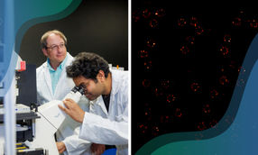
An optical laser can be used to align complex biomolecules for single-molecule imaging.
DESY-CFEL–CMI, Muhamed Amin
Laser-induced alignment utilizes the interaction between the anisotropic polarizability of the molecule and the electric field of a laser pulse. The laser pulse induces a transient electric dipole moment in the molecule, which then follows the electric field of the laser pulse. This forces the molecules to rotate into an orientation where the polarizability interaction is optimized, i.e., to align the most-polarizable axis of the molecule with the laser polarization, resulting in the geometric alignment of the molecules and thus fixing it in space.
In the current work, the researchers computationally demonstrated that nanoparticles and proteins can be strongly aligned using current standard laser technologies. Analyzing 150 000 proteins from the international protein databank (PDB), they showed that most proteins could be aligned using realistic experimental conditions. This enhances their visibility in single-particle diffraction experiments. The team published their findings in the Journal of the American Chemical Society.
The findings address a long-standing issue in single-particle imaging, where molecules are typically captured in random orientations, making 3D reconstruction difficult. The researchers also predict that cooling the molecules to cryogenic temperatures, a technique the group is actively implementing, enhances the degree of alignment and also reduces potential radiation damage, further refining the technique.
This breakthrough has significant implications for structural biology and nanotechnology, enabling scientists to visualize molecular structures with unprecedented detail, and could potentially revolutionize drug discovery, biomolecular research, and materials science. Future experiments will focus on integrating these laser techniques with XFEL imaging to achieve sub-nanometer resolution, bringing researchers closer to real-time visualization of molecular dynamics.
Original publication
Other news from the department science


These products might interest you

Eclipse by Wyatt Technology
FFF-MALS system for separation and characterization of macromolecules and nanoparticles
The latest and most innovative FFF system designed for highest usability, robustness and data quality

DynaPro Plate Reader III by Wyatt Technology
Screening of biopharmaceuticals and proteins with high-throughput dynamic light scattering (DLS)
Efficiently characterize your sample quality and stability from lead discovery to quality control