What our brain does when the mind is at rest
New insights into the brain’s default mode network

The brain’s default mode network (DMN) is a group of regions that become active when we are not engaged with our surroundings – for instance, when daydreaming. When solving tasks, however, this network is less active. Researchers at Forschungszentrum Jülich in Germany have now investigated the structure and function of this network by analysing brain tissue and applying advanced magnetic resonance imaging techniques. Their study revealed microstructural differences that influence how the DMN communicates with other regions of the brain. The findings have been published in the journal Nature Neuroscience.
The DMN includes regions such as the parahippocampus, precuneus, middle temporal lobe and parts of the frontal lobe – areas associated with memory, self-awareness and the processing of past experiences. It supports what is known as stimulus-independent thought – cognitive processes that are not triggered by external sensory stimuli. These include not only daydreams but also planning for the future or reflecting on the past as well as social situations and self-reflection. Moments of creative insight also often emerge during such inward-focused mental states.
However, the DMN also plays a role in more demanding cognitive tasks and, under certain circumstances, can be influenced by external stimuli. This led neuroscientists to a paradox: how can a network known for stimulus-independent thought also respond to sensory input? The new study offers an explanation. The DMN is not a uniform system, but rather consists of structurally distinct subregions. Some areas are closely linked to sensory brain regions and can be triggered by external cues such as sounds or smells. Others are more insulated and support introspective thinking that arises internally.
How the DMN processes information
The researchers from the Institute of Neuroscience and Medicine (divisions INM-1 and INM-7) found that the brain’s architecture determines not only its structure, but also how it functions – from simple perception to complex cognitive abilities. Their findings offer important insights into why certain thoughts are strongly influenced by sensory input – for instance, when a particular scent evokes a memory or a piece of music stirs an emotion – and how the DMN translates such stimuli into our inner world of thought.
The more clearly layered areas of the DMN resemble the architecture of sensory brain regions, which are responsible for processing inputs such as sight and sound. Like those regions, they consist of several distinctive cellular layers responsible for different types of information processing. Some layers receive signals from other brain areas, while others are responsible for relaying this information. In the DMN, this layered structure supports stronger connectivity with other brain regions and more efficient processing of sensory data.
This could explain why the DMN becomes active when external stimuli like smells or music evoke memories or emotions. The less strongly layered regions, by contrast, are more internally self-contained and less affected by the outside world. They support introspective thinking such as daydreaming or self-reflection – processes that arise from within.
Two approaches for new insights
To investigate the DMN’s structure and function in more detail, the research team combined two methods: in-depth histological analysis of post-mortem brain tissue and advanced magnetic resonance imaging of living participants.
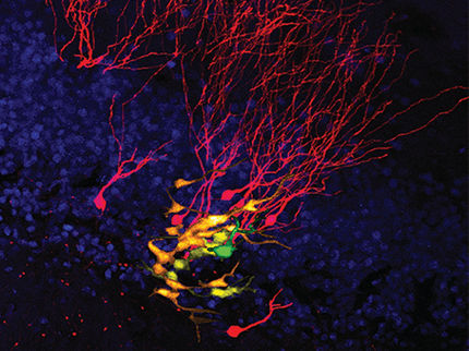
For the tissue analyses, thin slices of brain tissue were examined under the microscope. This revealed fine microstructural details – such as how neurons in different parts of the DMN are arranged and how they form into layers.
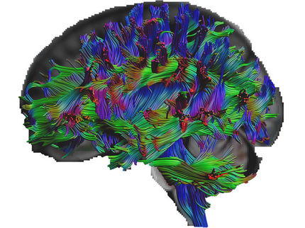
For the MRI scans in living participants, the researchers used a non-invasive technique to study brain structure and measure activity, without requiring any surgical procedure. This allowed them to observe which parts of the DMN are particularly active and how they interact with other brain areas.
Combining both methods proved to be the key. MRI alone cannot usually detect such fine structural detail. However, by comparing the scans with actual tissue samples, the researchers were able to identify patterns of nerve cell organisation that also play a role in the living brain. This allowed them to demonstrate that microstructural features – such as the layering of neurons – directly influence how information is processed and transmitted within the DMN. Many of the patterns observed in the tissue samples could also be confirmed in the brains of living individuals.
Original publication
Casey Paquola, Margaret Garber, Stefan Frässle, Jessica Royer, Yigu Zhou, Shahin Tavakol, Raul Rodriguez-Cruces, Donna Gift Cabalo, Sofie Valk, Simon B. Eickhoff, Daniel S. Margulies, Alan Evans, Katrin Amunts, Elizabeth Jefferies, Jonathan Smallwood, Boris C. Bernhardt; "The architecture of the human default mode network explored through cytoarchitecture, wiring and signal flow"; Nature Neuroscience, Volume 28, 2025-1-28
Other news from the department science