What does "resolution" mean here? Microscopy puzzle solved
New microscopy method can identify molecules: The question of resolution proved to be a difficult puzzle

When judging the quality of a microscope, the crucial question is: How large are the smallest structures that can just be made visible with it? How close can two objects come to each other before they can no longer be seen as two separate objects, but blur into a single image blob?
With conventional light microscopes, this can be calculated using relatively simple formulas. However, complicated microscopy techniques are now used in many areas where this question is much more difficult to answer.
One of these is atomic force microscopy-infrared spectroscopy (AFM-IR), which can be used to map the distribution of chemical substances. This method can be used to identify and visualize proteins in a cell, for example. However, it has often been unclear how well this method works in which situation. The resolution of the method varies and depends in a complicated way on many different effects. The TU Vienna has now succeeded in describing these effects and calculating the resolution of such microscopes. What could previously only be determined by trial and error can now be reliably predicted.
Atomic force microscopes and infrared radiation
The AFM-IR microscopy technique has been the subject of research at TU Wien for several years. It combines atomic force microscopy (AFM) with infrared spectroscopy (IR).
Infrared radiation can be used to detect large molecules such as proteins: Different molecules react to different infrared wavelengths. By measuring at many different infrared wavelengths, a so-called infrared spectrum is obtained - something like the fingerprint of a molecule. This spectrum can be used to identify which molecule you are dealing with.
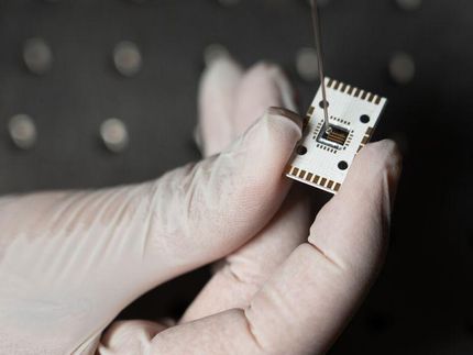
"However, you still don't know exactly where this molecule is located," says Prof. Georg Ramer from the Institute of Chemical Technologies and Analytics at TU Wien. However, this infrared method can be combined with an atomic force microscope. This involves scanning the surface of the sample with a very fine tip. If a molecule that is currently absorbing infrared radiation is located at a certain point, this leads to localized heating at precisely this point. The sample expands a little and this can be measured with the atomic force microscope. You then not only know which molecule it is, but also exactly where it is located.
The exact resolution? A mystery
"Many researchers and companies use this method with success because it can tell you with very high resolution where which molecules are located. Until now, however, it has been something of a dirty secret," says Georg Ramer. "Nobody could say how high the spatial resolution of the technology is. The answers you find in the literature - 10 nanometers or even 100 nanometers - are rarely really well-founded, but rather guesses." The method does not always work equally well; it varies from sample to sample.
This is a problem, because if you don't know the resolution, you can't say which applications the method can be used for. You may be carrying out experiments for which this technique is not actually suitable.
"We took a closer look at this and carried out experiments as well as developing computational models and computer simulations," says Yide Zhang, one of the two doctoral students working on the project. "We can now finally explain exactly why this strange effect occurs, where the resolution is sometimes better and sometimes worse."
When a molecule on the sample absorbs infrared light and heats up, this does not always lead to the same measured expansion. This expansion also depends on how quickly the heat is dissipated and how much material is between the molecule and the tip of the instrument. The new computer model can be used to calculate how strongly each sample reacts to this heat effect, and in which specific cases this effect should be visible and in which it should not.
Learning more about the sample than ever before
"Our results can now be used to decide in advance whether a particular experiment with the method makes sense at all," says Georg Ramer. "And that's not all: our work also allows us to interpret experiments more correctly and optimize sensitivity and resolution." Until now, for example, the sample was usually viewed as a two-dimensional surface. With the new findings, however, statements can now also be made about the third dimension: It is now possible to create a 3D image of the sample on a nanometer scale.
Note: This article has been translated using a computer system without human intervention. LUMITOS offers these automatic translations to present a wider range of current news. Since this article has been translated with automatic translation, it is possible that it contains errors in vocabulary, syntax or grammar. The original article in German can be found here.
Original publication
Yide Zhang, Ufuk Yilmaz, Gustavo Vinicius Bassi Lukasievicz, Liam O’Faolain, Bernhard Lendl, Georg Ramer; "An analytical model of label-free nanoscale chemical imaging reveals avenues toward improved spatial resolution and sensitivity"; Proceedings of the National Academy of Sciences, Volume 122, 2025-1-24

Other news from the department science


Most read news


More news from our other portals


See the theme worlds for related content
Topic World Spectroscopy
Investigation with spectroscopy gives us unique insights into the composition and structure of materials. From UV-Vis spectroscopy to infrared and Raman spectroscopy to fluorescence and atomic absorption spectroscopy, spectroscopy offers us a wide range of analytical techniques to precisely characterize substances. Immerse yourself in the fascinating world of spectroscopy!