Nerve cells of blind mice retain their visual function
Good news for retina implants
The retina is often referred to as an “outpost of the brain” – after all, important steps in visual signal processing do not take place in the cerebrum, but in the nerve cells in the eye. When light falls on the retina, sensor cells become active and send electrical signals to layers of nerve cells located directly behind them. From there, signals are passed on to the brain.

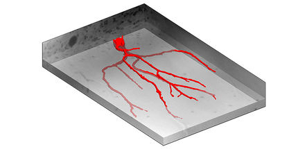
Printed micro-electrode arrays and patch-clamp setup used to record neuronal activity from ex-vivo mouse retina
Technische Universität Wien
However, it was previously unclear exactly how the signals from the retina are processed by the nerve cells. Experiments at TU Wien (Vienna) have now shown that the nerve cells of the retina (the so-called retinal ganglion cells) can take on different roles and thus fulfil individually different tasks for vision. They retain this ability even when parts of the retina degenerate – which is good news for the restoration of sight in blind people using electronic retinal implants, for example.
Different cells, different signalling patterns
“When light falls onto the photoreceptors of the retina, electrical signals are generated in the nerve cells behind them,” says Paul Werginz from the Institute of Biomedical Electronics at TU Wien. “But not all nerve cells produce the same sequence of signals.” When light is switched on or off, certain types of nerve cells always become active. However, in some cells the frequency of the signals quickly decreases, while other cells remain at a comparatively high level of activity and continue to emit a strong electrical signal.
It has been unclear what causes these different activity patterns. After all, cells of the same type should be expected to behave similarly. “The question for us was: if the retinal ganglion cells behave differently, is it because they are integrated into different biological circuits and therefore receive different input signals? Or is there an intrinsic difference based on biophysical principles that causes these cells to produce different signals, even if they receive identical inputs?” says Paul Werginz. “In the second case, each ganglion cell type could be assigned its own component ID, so to speak.”
Electrical impulses instead of light
To test this, the researchers used explanted retinas from mice in which the entire neuronal network is kept functional for several hours. The activity of the retinal ganglion cells can then be stimulated in two different ways: Either by irradiating the retina with light and then investigating how the ganglion cells react, or by directly stimulating the ganglion cells using electric current. The direct injection of electric current makes it possible to investigate the properties of the neurons even without involving the cells that usually provide them with input.
“We found that when we directly stimulate the cells with electric current, they show a signalling pattern very similar to the one they produce when exposed to light,” says Paul Werginz. “Ganglion cells that show an increased activity pattern for a longer time period when exposed to light also do so when electrically stimulated.”
This means that the difference between the signalling patterns of these cells is not only due to the fact that they receive different input in the retinal circuitry – the tendency to produce longer or shorter signalling sequences is an intrinsic property of the cells.
“This is astonishing but is likely to be very important for signal processing and vision,” believes Paul Werginz. “These differences between the cell types probably arise very early on, during the developmental phase of the retina.”
Stable differences - even with blindness
An important question remains: if these are intrinsic properties of the cells, do these properties remain stable even if the cells lose their original function – for example, if the retina's photosensors no longer work? One might assume that the behaviour of the cells should change in this case. After all, it has often been observed that nerve cells which are no longer needed are reorganized inside the brain. If one loses a finger, for example, the nerve cells that were responsible for the sensory signals from this finger do not simply remain inactive, they become rewired and re-used for other purposes.
However, this is different for retinal ganglion cells: “We examined the cells of mice that had been blind for 200 days, and their retinal ganglion cells still showed exactly the same properties: some could be made to be active for a short time with electrical input, others for longer,” says Paul Werginz. The cells therefore retain their intrinsic ability to deliver certain signals.
This is good news for the development of retinal implants that use electrical stimulation via thousands of electrodes to replace the lost photoreceptors in blind patients, says Paul Werginz: “If there are stable differences between different cell types, then the existing ganglion cells can be utilised even after blindness and better stimulation strategies can be developed for them in the future.”


