Intricate processes in photosynthesis decoded using advanced electron microscopy technique
This enables breakthroughs in multiple areas of biological and chemical research

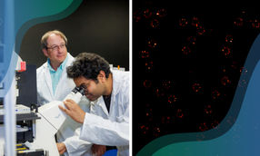
Using cryo-electron microscopy a team of scientists from Humboldt-Universität zu Berlin (HU), the Swedish universities of Umeå and Uppsala and the University of Potsdam has succeeded in visualising atomic structures at an unprecedented resolution at the nanometre level underlying the process of photosynthesis. For the study, which was published in the journal Science, the team specifically studied the protein structure known as Photosystem II, in which the first step of photosynthesis takes place: Light is absorbed and used as an energy source to drive the splitting of water molecules into oxygen, protons and electrons.
A decisive step towards understanding photosynthesis
The high-resolution visualisation provides new insights into the interactions of hydrogens within photosystem II, which are crucial for the reaction driven by light energy. The team, led by Dr Rana Hussein and Prof Dr Athina Zouni from the Department of Biology at HU, Prof Dr Wolfgang Schröder from Umeå University and Prof Dr Johannes Messinger from Uppsala University, has thus taken a significant step in understanding the complex processes of photosynthesis.
"By using cryo-electron microscopy, we can now observe the locations of hydrogens in photosystem II," says Prof Dr Athina Zouni. "This detailed view is crucial for understanding the process by which oxygen-evolving organisms convert light energy into chemical energy - a process that is fundamental to life on Earth."
Prof Dr Holger Dobbek elaborates on the most important results of the study: "We use cryo-electron microscopy to show photosystem II with better resolution. This enabled us to detect hydrogens in several amino acid residues in the reaction centre sites, providing new information on the transfer of electrons and protons in photosystem II. Our research reveals the sequence of events leading to the second protonation of a mobile plastoquinone B. This profoundly renews our understanding of the electron transport chain in photosynthesis."
Research method reaches far beyond the field of photosynthesis
Dr Rana Hussein explains: "The innovative approach used in this study to determine the positions of protons and hydrogens is essential for understanding photosystem II and has a broad spectrum of applications. It can be applied to study various proteins to uncover mechanisms regarding hydrogens. This enables breakthroughs in multiple areas of biological and chemical research. Thus, the cryo-EM method used in this study has implications beyond photosynthesis ".
In cryo-electron microscopy, protein complexes are cooled down to very low temperatures of up to -260 °C within fractions of a second. This shock freezing prevents the formation of ice crystals so that molecules keep their natural form. In the future, the visualisation of hydrogens could contribute to understanding other fundamental biochemical reactions, such as enzyme mechanisms, protein-ligand interactions or the dynamics of membrane proteins.
Original publication
Other news from the department science