It is in our genes – and how our genome folds in 3D
New findings could help in the prevention and treatment of diseases
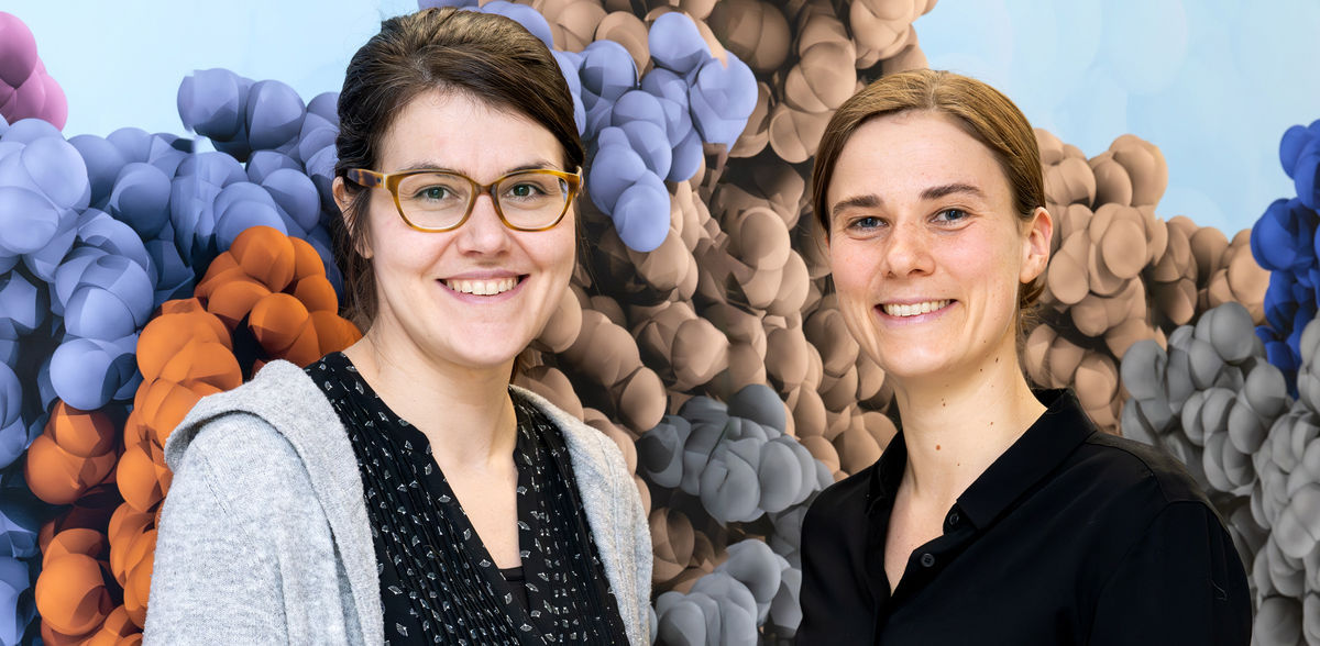
Whether we stay healthy or become seriously ill is determined by our genes. But also, the folding of our genome has a significant influence on this, as the 3D genome organization regulates which genes are switched on and off. Researchers led by Marieke Oudelaar and Elisa Oberbeckmann at the Max Planck Institute (MPI) for Multidisciplinary Sciences have now succeeded in recreating the 3D folding of the yeast genome in the laboratory and deciphering the underlying mechanisms.
Our body consists of hundreds of different cell types that are specialized for various tasks: They fight pathogens, transport oxygen, produce insulin, or make us think. All cell types contain the same blueprint, which is encoded in the genes in our DNA. However, only the genes that the respective cell needs for its task are activated.
The DNA of a cell in our body, our genome, would be about two meters long if stretched out. Nevertheless, the DNA fits into a tiny cell nucleus with a diameter of around one-thousandth of a millimeter. It is therefore compactly packaged. Certain proteins, the histones, wind up the DNA in sections like on a cable drum. This histone-DNA complex is called a nucleosome. The nucleosomes are separated from each other by nucleosome-free DNA sections, similar to how knots are strung together in a thread. The genome packed into nucleosomes is called chromatin.
Accessible and activatable genes
“The 3D structure of the genome influences the activity of the genes. By folding, regulatory DNA sections come into contact with the right genes at the right time and switch them on or off,” explains Marieke Oudelaar, who heads a Lise Meitner research group at the institute. If gene activation is disrupted, this can cause various diseases in humans, such as cancer.
It is still a challenge for scientists to investigate the 3D structure of the DNA: “In living cells, it is difficult to uncover which proteins and processes are involved in 3D genome folding. The underlying biochemical networks are difficult to disentangle, and the proteins involved often have several functions that are difficult to separate,” explains Elisa Oberbeckmann, project group leader at the MPI and first author of the study now published in the journal Nature Genetics.
Chromatin recreated in the lab
The researchers and their team have now achieved an important methodological advance: They succeeded in reproducing chromatin from yeast in the laboratory and measuring its 3D structure. “This offers the great advantage that the folding process and the proteins involved can be studied in isolation,” says Oberbeckmann. Using biochemical and genetic experiments as well as computer simulations, they were then able to decipher how chromatin folds into specific 3D structures called chromatin domains.
When the researchers added regulators that remodel chromatin in the laboratory experiment, the artificially generated chromatin formed 3D structures very similar to the chromatin in yeast. “To our surprise, it folded independently as soon as cellular regulators brought a certain regularity to the chromatin. The regular arrangement of the nucleosomes through chromatin remodeling appears to play a much more central role in the 3D folding of the genome than previously thought,” explains Oudelaar.
The team could show that long nucleosome-free sections in the chromatin form the boundaries between the individual domains. “The length of these sections directly influences whether neighboring domains can interact with each other. Longer nucleosome-free regions are very rigid and thus prevent interactions between nucleosomes from two adjacent domains,” explains Kimberly Quililan, a student who played a key role in the laboratory experiments and analyses.
Not only relevant for yeast
The insights gained by the researchers are also relevant beyond the yeast model system: Similar mechanisms could play a role in the 3D organization of the human genome. In the future, the results of the Göttingen team could help to better identify possible causes of diseases that are based on a disruption of gene regulation. “If we understand how genes become accessible in the 3D genome and are activated, this could also be a useful starting point for the prevention and treatment of such diseases,” says Oudelaar.


