Researchers develop new marker for cancer cell motility
Physics of Cancer finds first potential application in oncology – More accurate prognosis of how breast tumours spread
Researchers led by Leipzig University have found a ground-breaking application in oncology for the scientific field of Physics of cancer. This is a milestone for the new research field, proving its clinical relevance for the first time. Based on tissue and cell mechanics and using machine learning, the team developed a marker for cancer cell motility in digital pathology. The marker delivers new information about breast tumours that will improve the ability to predict the risk of metastasis, even after a decade has passed. The researchers have just published their new findings in the journal Physical Review X.

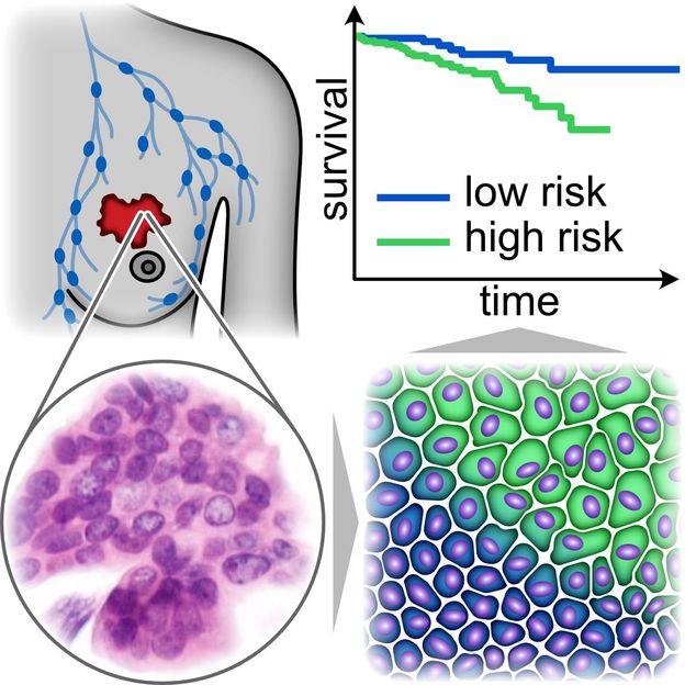
Two aggregate states of cancer cell clusters. In blue: densely packed, more rounded cancer cells jam together and cannot move, reducing the risk of metastasis. In green: more elongated, loosely packed cancer cells can push past each other.
Dr. Hans Kubitschke
In a retrospective study of 1,380 female breast cancer patients, conducted in close collaboration with Professor Axel Niendorf from the Pathologie Hamburg-West institute, doctoral researcher Pablo Gottheil from the research group led by Professor Josef Alfons Käs at Leipzig University found that a collective transition of cancer cells to motility, known in specialist circles as “unjamming”, significantly increases the risk of distant metastases. Distant metastases occur when cancer cells spread to other organs. “This process plays a crucial role in the aggressiveness of cancer and could be an important additional prognostic factor for the risk of a tumour spreading,” says Professor Josef Alfons Käs, head of the Soft Matter Physics Division at Leipzig University.
In the primary tumour, the cancer cells form clusters where they are packed so tightly that they “jam” together and cannot move. As the tumour grows, a collective transition to motility occurs, allowing the cancer cells to leave the tumour and spread. Here the cancer cells take on an elongated shape that allows them to push past each other. The histological sections used to diagnose breast cancer are static images that do not show how cells move. “What we have now found is that we can identify motile cancer cells in these histological images on the basis of their elongated shape and lower density. We have identified a first marker that allows the detection of motile cancer cells in histological tumour sections. These cells are able to spread and disperse. We were able to show that a high number of such motile cells in a tumour sample significantly increases the risk of metastasis,” explains biophysicist Käs.
Reducing over- and under-treatment of patients
“More precise diagnostics could significantly improve our ability to treat breast cancer. There are many different, differentiated approaches to treatment. However, since current diagnostics cannot accurately predict disease progression, patients are over- and under-treated,” says Käs. Predicting whether metastases will form in the body is an important aspect of prognosis. Diseased lymph nodes in the armpit pose a high risk. However, about 30 per cent of women with affected lymph nodes will not develop distant metastases, whereas the tumour may spread within the body in 30 per cent of women whose lymph nodes are not affected.
According to the underlying study, the new prognostic marker developed by Käs and his colleagues has a clinically relevant predictive power that is comparable to the lymph node status previously used in diagnostics. The researchers believe that the two diagnostic criteria are complementary in their predictive power and can correct each other’s false predictions. This has the potential to reduce the number of women who are over- or under-treated. Most importantly, the new marker allows for an earlier prognosis because it can be used to make a prediction before the cancer cells have left the tumour. This means that a prognosis can also be made for patients in the early stages of the disease, when lymph node status is not yet useful. The researchers’ new approach could therefore be important for the early detection of particularly aggressive tumours.
Procedure also applicable to other tumour types
These results could only be achieved by working closely with medical professionals. According to Käs, the study would not have been possible without the large number of digital histological sections of breast tumours provided by Professor Niendorf. The Department of Gynaecology at the University of Leipzig Medical Center, headed by Professor Bahriye Aktas, provided vital breast tumour explants. In this way, the biophysicists were able to confirm that the morphological criteria for “unjamming” do in fact apply to motile cancer cells. Professor Markus Löffler from the Institute for Medical Informatics, Statistics and Epidemiology at Leipzig University and his team supported the project with clinical evaluation and interpretation.
“I am thrilled about these exciting results! With this evaluation of unjamming, we will have another useful prognostic marker to add to our current diagnostic toolbox. But before the marker can be used in clinical practice, further prospective studies are needed,” says Professor Aktas.
Since the method does not rely on specific molecules in the tumour, it could also be used for other tumour types. According to the researchers, the procedure could therefore be applied to 92 per cent of all cancer patients. In addition, “unjamming” can be an important pathological factor in other diseases, such as asthma.
With more than two million cases worldwide each year, breast cancer is by far the most common cancer in women. In 2018, more than 600,000 women with breast cancer died, primarily due to the systemic, invasive nature of the disease.
Original publication
Pablo Gottheil, Jürgen Lippoldt, Steffen Grosser, Frédéric Renner, Mohamad Saibah, Dimitrij Tschodu, Anne-Kathrin Poßögel, Anne-Sophie Wegscheider, Bernhard Ulm, Kay Friedrichs, Christoph Lindner, Christoph Engel, Markus Löffler, Benjamin Wolf, Michael Höckel, Bahriye Aktas, Hans Kubitschke, Axel Niendorf, and Josef A. Käs: "State of Cell Unjamming Correlates with Distant Metastasis in Cancer Patients"; Physical Review X; 2023


