Brain research with organoids
Scientists develop effective method to genetically modify brain organoids
Primates are among the most intelligent creatures with distinct cognitive abilities. Their brains are relatively large in relation to their body stature and have a complex structure. However, how the brain has developed over the course of evolution and which genes are responsible for the high cognitive abilities is still largely unclear. The better our understanding of the role of genes in brain development, the more likely it will be that we will be able to develop treatments for serious brain diseases. Researchers are approaching these questions by knocking out or activating individual genes and thus drawing conclusions about their role in brain development. To avoid animal experiments as far as possible, brain organoids are used as an alternative. These three-dimensional cell structures, which are only a few millimeters in size, reflect different stages of brain development and can be genetically modified. However, such modifications are usually very complex, lengthy and costly. Researchers at the German Primate Center (DPZ) – Leibniz Institute for Primate Research in Göttingen have now succeeded in genetically manipulating brain organoids quickly and effectively. The procedure requires only a few days instead of the usual several months and can be used for organoids of different primate species. The brain organoids thus enable comparative studies of the function of genes at early stages of brain development in primates and help to better understand neurological diseases (Jove Journal).

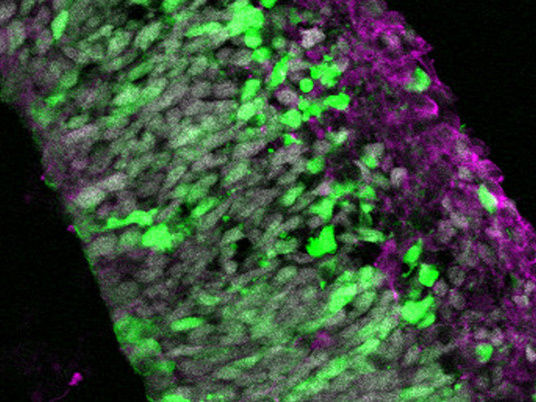
Section of an electroporated brain organoid of a common marmoset. Green: electroporated cells that glow green due to the green fluorescent protein; magenta: neurons; gray: nuclei.
Lidiia Tynianskaia
Brain organoids are grown in the laboratory from so-called induced pluripotent stem cells. These cells are usually derived from skin or blood cells that are "reprogrammed" beforehand. That is, they are modified so that they regress to stem cells and can then differentiate into any other cell type, such as neurons.
"We are particularly interested in the genetic factors underlying brain development in primates," explains Michael Heide, head of the Junior Research Group Brain Development and Evolution at DPZ and author of the study. "The brain organoids allow us to reproduce these processes in the Petri dish. To do that, however, we need to genetically modify them."
Until now, these procedures were sometimes very labor-intensive and took several months. The team of researchers led by Michael Heide has now developed a method that allows brain organoids to be genetically manipulated quickly and cost-effectively.
"We use microinjection and electroporation for our method," Heide explains. "In this process, genetic material is injected into the organoids with a very thin cannula and introduced into the cells with the help of a small electrical pulse. It takes only a few minutes, and the brain organoids can be analyzed after a few days."
Plasmids are used to insert the genetic material. These are circular pieces of DNA that contain the gene of interest. In the feasibility study, the researchers used the green fluorescent protein (GFP) gene for this purpose. Successfully modified cells in brain organoids thus glow green under fluorescent light.
"The method is equally suitable for brain organoids from humans, chimpanzees, rhesus macaques and common marmosets," Heide summarizes. "This allows us to perform comparative studies on physiological and evolutionary brain development in primates and is also an effective tool to simulate genetically caused neurological malformations without having to use monkeys in animal experiments."


