Neuroscientists illuminate how brain cells deep in the cortex operate in freely moving mice
This new miniature microscope is a game changer for exploring the link between neural activity and complex animal behavior
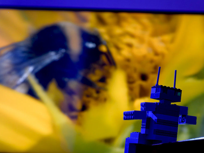
How can we see what neurons deep in the cortex are doing during behavior? Researchers at the Max Planck Institute for the Neurobiology of Behavior - caesar (MPINB) have developed a miniature microscope small enough to be carried on the head of a freely moving mouse and capable of measuring neuronal activity in all cortical layers, even the deepest ones. The two-gram microscope can be controlled remotely, which minimizes the need to handle the animal. The microscope also incorporates new technology enabling imaging in lit environments, something that all comparable microscopes struggled to do. Neuronal activity can now be imaged from all cortical layers in the freely moving mouse during the full range of the animal’s behaviors. This new microscope is a game changer for exploring the link between neural activity and complex animal behavior.

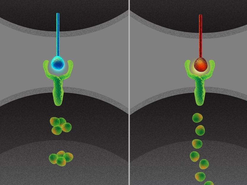
Illustration für die Publikation "A three-photon head-mounted microscope for imaging all layers of visual cortex in freely moving mice"
Julia Kuhl
How does the brain produce behavior? Studying the brain during behavior is most informative when the animal is free to interact with its environment in any way it chooses. This requires small head-mounted devices that provide access to the brain, but that do not interfere with the animal’s behavior. “We are interested in how freely behaving animals use vision to make decisions in their everyday life, and as many of the brain cells that are thought to be involved in this process are located deep in the visual cortex, we made a very light-weight head-mounted microscope that can measure activity from these neurons but does not interfere with the animal’s behavior. We have made a large step towards finally imaging brain activity deep in the cortex of animals performing natural visual-based behaviors” said Jason Kerr, director of the Department of Behavior and Brain Organization.
In the study that is published on November 28, 2022 in Nature Methods, the researchers developed a miniature 2-gram three-photon excitation microscope which delivers a number of firsts. For the first time, it is now possible to image neural activity on a single-cell level from all cortical layers without having to interfere with the animal’s behavior, made possible by a remote focusing mechanism. Its modular design also offers a high-resolution configuration which allows for functional recordings from neuronal somata and dendrites. Another important feature is that, due to a modified detector system, the microscope can be used in fully lit conditions. “The robustness of our new miniature microscope to ambient light allows us to image the brain’s activity while the animal is having access to its full sensory repertoire. We can now study visually guided natural behavior like prey hunting and predator avoidance” says Alexandr Klioutchnikov, first author of the study. To confirm range and stability of the new miniature three-photon microscope, the team imaged from the deep cortical layers 4 (L4) and 6 (L6) while mice were freely exploring an arena. It turned out that L4 and L6 neurons were differentially modulated depending on the environmental light.
As the microscope can easily be mounted again at the exact same position, the same neuronal populations can be imaged in follow-up sessions spread over days. This opens up the possibility to monitor changes in the brain’s activity, for example while the animal is learning.
Original publication
Other news from the department science


These products might interest you
alpha300 R by WITec
3D Raman microscopes with unequalled speed, sensitivity and resolution
Visualize and characterize every chemical detail

JEOL CRYO ARM by JEOL
Cryo-TEM: Fast and stable data acquisition for biosamples
Increased efficiency in structural biology with automated sample loading system
FLUOVIEW FV4000 by EVIDENT
Revolutionary imaging with FLUOVIEW FV4000: Confocal laser scanning
Utilise AI-powered image processing and innovative detector technology

Most read news


More news from our other portals


Last viewed contents
Neo-Darwinism
Griffith's_experiment
Artificial_pacemaker
Peptide_synthesis
Antibody
Radioresistance
Cells communicate in a dynamic code - Important intercellular communication encodes and transmits more than thought
Evolutionary_history_of_plants
Ellis-van_Creveld_syndrome
Conservation_Commons
Lactic_acid_fermentation