Candida albicans digs transcellular tunnels
Generally harmless, the fungus Candida albicans is found in the microbiota of a large number of individuals. However, it can become pathogenic in certain situations, even dangerous for fragile people. In these people, it can indeed make its way through the intestinal wall and enter the bloodstream to invade the whole body. A team of scientists trying to understand the details of the mechanisms involved in these invasions has just discovered a curious phenomenon: the microorganism is capable of digging transcellular tunnels...
Candida albicans is a fungus - and more precisely a yeast - naturally present in the oral, vaginal and digestive mucosa of humans. It is part of the microbiota of many people, along with countless other microorganisms. Most often its presence does not interfere with our health, even if its proliferation can lead to local infections called "thrush" or "candidiasis". However, in certain fragile people, especially when they are immunocompromised, this yeast can become seriously pathogenic. It then takes an invasive filamentous form, called "hyphae", which colonizes the intestinal walls, reaches the bloodstream and leads to a generalized infection.
Work has already been done on the mechanisms that allow Candida albicans to evolve from its classic round form to the filamentous form capable of infiltrating tissues. Thus, the major early stages of infection are already fairly well known: the yeast attaches to cells of the intestinal wall via adhesion molecules, then the hypha forms in response to environmental factors and invades adjacent tissue. But paradoxically, all this happens initially without damaging host cells or breaking their membranes: a silent progression that intrigued Allon Weiner, a researcher at the Center for Immunology and Infectious Diseases in Paris. " How can yeast enter cells without damaging them? That was a mystery until now," he says.
Video and electron microscopy to the rescue
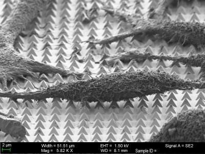
To study this phenomenon, his team used video-microscopy and 3D volume electron microscopy. Applied to living cells, video microscopy allows the observation of invasion in real time, while electron microscopy allows the visualization of events at the nanoscale within fixed cells. The researchers worked with different cell lines brought into contact with C.albicans in vitro, including intestinal wall cells (enterocytes) that express a fluorescent version of galectin-3. This protein is recruited in case of damage to the cell membrane, and then involved in its repair. Transformed into a fluorescent marker, it makes it easy to identify cells that have been damaged by C. albicans.
This is how the researchers observed a novel invasion process, which allows the silent progression of hyphae through cells without destroying them, via transcellular tunnels. How do they do this? By pushing back the membranes of the host cells, which then form the walls of these tunnels: " It's as if your finger were the hypha and you pushed it into several superimposed balloons, until you had passed through all of them without bursting them," Allon Weiner illustrates. These tunnels are also wider in some places where there is glycogen, which is known to be a nutrient for C. albicans. " This observation deserves to be further explored, as it suggests the existence of glycogen refueling zones for the yeast within these tunnels," says the researcher.
The formation of these tunnels takes place over several hours, until the fungus begins to damage the cells. It is only between 9 and 24 hours after the beginning of the infection that cells start to be destroyed. This is problematic from an immune standpoint," says Allon Weiner. Since the progression of the fungus through the intestinal wall is silent in the first few hours, there is no signal to the immune system to block the infection at this early stage. " Other work confirms this: the C. albicans infection produces only low levels of pro-inflammatory cytokines during its first few hours. But Allon Weiner believes that by further unraveling these mechanisms, it should be possible to learn how to counteract this mechanism to prevent severe infections. He is already at work...
Note: This article has been translated using a computer system without human intervention. LUMITOS offers these automatic translations to present a wider range of current news. Since this article has been translated with automatic translation, it is possible that it contains errors in vocabulary, syntax or grammar. The original article in French can be found here.


