Study of skin biopsies offers potential as new diagnostic marker for Amyotrophic Lateral Sclerosis
The discovery of this biomarker detectable in small skin samples can be very useful for cases in which diagnosis is difficult
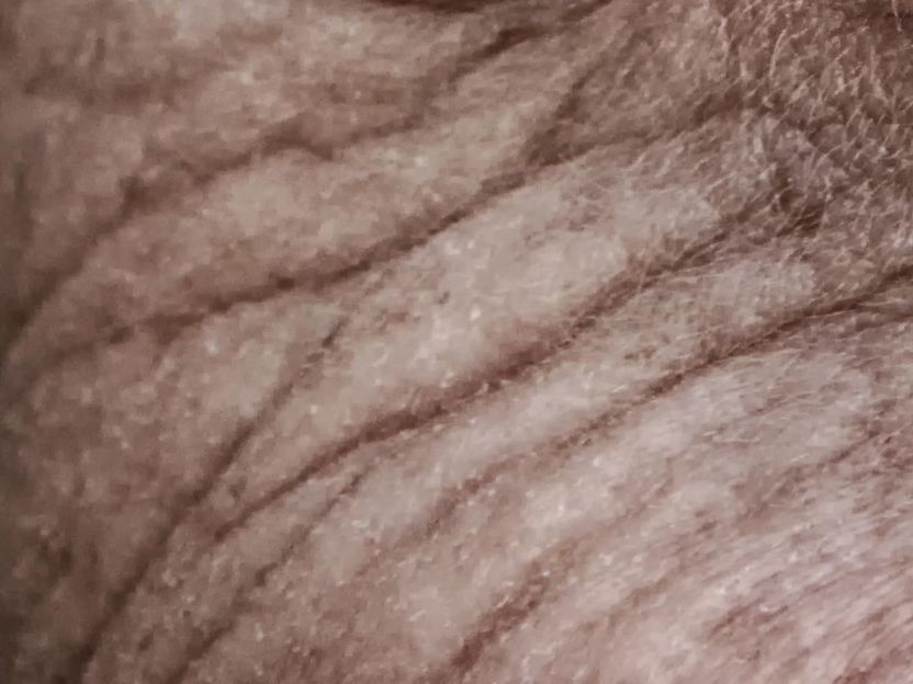
Barcelona (UAB) and of the Functional Unit for amyotrophic lateral sclerosis at the Hospital del Mar have identified the presence of an ALS biomarker in the skin of the patients. This specific protein, TDP-43, is present outside the nucleus of skin cells of patients. The presence in the skin of an elevated number of cells with this abnormality allows to predict whether the patient has the disease or not, apart from how far it has progressed.

analogicus from Pixabay
The research, published in the journal Cells, was led by Dr Xavier Navarro and Dr Mireia Herrando-Grabulosa, both from the Neuroplasticity and Regeneration Group of the Department of Cell Biology, Physiology and Immunology and the Institut de Neurociències of the UAB, and by Dr Miguel Ángel Rubio, coordinator of the ALS Functional Unit at Hospital del Mar. They analyzed skin samples of patients with ALS from this centre and the Bellvitge University Hospital. The results were compared to those of ten healthy subjects and ten other patients suffering from other neurodegenerative diseases with chronic neuroinflammation component. The researchers studied the presence of TDP-43 protein in skin cells, since this protein is found in all of the body cells and is basic for developing their functions, outside the cell nucleus, i.e. in the cytoplasm. It is known that its presence outside the nucleus in the motor neurons of the brain and the spinal cord is seen in 97% of ALS patients.
More common in ALS patients
The researchers were able to determine that the TDP-43 protein had also abandoned the cell nucleus in a large amount of skin cells of the ALS patients analyzed, using small skin biopsies. The same was not found in the samples of the subjects in the control groups. “In the skin layers analyzed, the ALS patients presented more fibroblasts, which are basic tissue cells in the dermis, with the mark of the disease which is normally seen in the spinal cord and the motor cortex, when compared to the members of the healthy control group and the control group with other pathologies”, Dr Rubio points out. In addition, skin samples from ALS patients were taken one year later, thus certifying that this situation continues to exist to the same extent, regardless of the evolution and progression of the disease.
Specifically, this anomaly is seen in one of every four skin tissue cells (in 24.1% of fibroblast cells) of ALS patients, while it is almost non-detectable in healthy individuals or in those with other neurodegenerative diseases. As Dr Rubio highlights, “we have a biomarker that works as a fingerprint of the disorder in the nervous system, and we also confirmed that it can be found in the skin. Moreover, we can quantify and determine the theoretical cutoff point to be able to give a diagnosis in specific cases”.
“It is probable that this marker, in pre-symptomatic stages, before having any initial motor manifestations, could already be present and that is why it may be relevant for a diagnosis”, explains Dr Rubio. He also made it clear that presently, this tool can help in cases of difficult diagnosis or in people with a family history of ALS who present mutations in the genes that can cause the disease. In any case, further studies are needed, with more patients, to certify that this new biomarker can be used to speed up the diagnosis of the disease.
Original publication
Other news from the department science
Most read news
More news from our other portals
See the theme worlds for related content
Topic world Diagnostics
Diagnostics is at the heart of modern medicine and forms a crucial interface between research and patient care in the biotech and pharmaceutical industries. It not only enables early detection and monitoring of disease, but also plays a central role in individualized medicine by enabling targeted therapies based on an individual's genetic and molecular signature.


