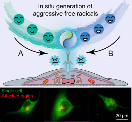
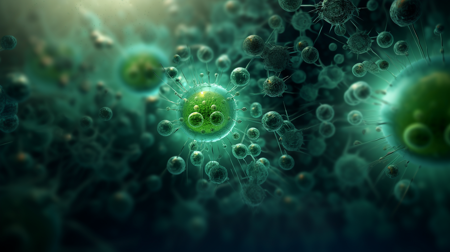
Radical Attack on Live Cells
Stimulation of tiny areas on cellular surfaces with free radicals using a microfluidic probe
Is there a way to chemically manipulate small, confined areas on cellular surfaces? Scientists have developed a microfluidic probe to send a flow of free radicals on live cells and track the outcome using fluorescence imaging. As outlined in the journal Angewandte Chemie, this approach makes it possible for the first time to generate a reaction zone of free radicals with controlled size and concentration for subcellular research.
© Wiley-VCH
Free radicals are important stimulants for cells. When live cells are exposed to radicals, they develop intense reactions that can lead to cell injury or even death. Many anticancer drugs are based on the action of free radicals sending cancer cells to death.
However, scientists find it difficult to perform research on the reactions of live cells to radicals in a truly controlled way. Free radicals are unstable and react with their environment before reaching their targets. A team of scientists led by Jin-Ming Lin from Tsinghua University, Beijing, has now developed a microfluidic approach to continuously generate a flow of free radicals for subcellular manipulation.
To make the radicals, the researchers chose a microfluidic two-component system. In this setup, one microchannel harbored a solution of enzymes able to cleave hydrogen peroxide. Another channel contained a solution of hydrogen peroxide and an organic dye. Both channels were immersed with their ends in a nutrient solution where a live cell was placed just below the channel ends. A third channel with an upward flow ensured that the fluids leaving the microchannel ends would meet in the middle position, forming a confined reaction zone.
According to the authors, this setup ensured that the reaction zone had the size of only a few micrometers. In this zone, the enzyme horseradish peroxidase would react with the hydrogen peroxide to form reactive enzyme intermediates, which then reacted with the organic dye to give an organic radical. Immediately after their generation, the dye radicals would then attack the cell placed directly below the reaction zone.
After tens of seconds of component flow and radical attack, the researchers observed that a tiny spot emitting bright red fluorescence had emerged on the cellular membrane. Tracking this spot over time, the researchers found it slowly wandered around on the cellular surface.
The authors say that the tiny fluorescent spot and its movement highlight the ability of the microfluidic method to manipulate small subareas on the cell surface. “By contrast with lipophilic tracers, which stain the whole cell, it is convincing that the free radicals generated only attack the target subcellular region of the single cell,” they argue.
One particular application fascinates the authors: they envision using the microfluidic probe as a “pen” for cells. “This will enable us to directly write text or draw graphics on single cells for personalized cell marking or artwork,” they explain.
Original publication
Other news from the department science


Most read news


More news from our other portals


See the theme worlds for related content
Topic world Fluorescence microscopy
Fluorescence microscopy has revolutionized life sciences, biotechnology and pharmaceuticals. With its ability to visualize specific molecules and structures in cells and tissues through fluorescent markers, it offers unique insights at the molecular and cellular level. With its high sensitivity and resolution, fluorescence microscopy facilitates the understanding of complex biological processes and drives innovation in therapy and diagnostics.
Topic World Cell Analysis
Cell analyse advanced method allows us to explore and understand cells in their many facets. From single cell analysis to flow cytometry and imaging technology, cell analysis provides us with valuable insights into the structure, function and interaction of cells. Whether in medicine, biological research or pharmacology, cell analysis is revolutionizing our understanding of disease, development and treatment options.