New microscopy analysis allows discovery of central adhesion complex
Researchers develop a new method for quantitative single-molecule colocalization analysis
Cells of organisms are organized in subcellular compartments that consist of many individual molecules. How these single proteins are organized on the molecular level remains unclear, because suitable analytical methods are still missing. Researchers at the University of Münster together with colleagues from the Max Planck Institute of Biochemistry (Munich) have established a new technique that enables quantifying molecular densities and nanoscale organizations of individual proteins inside cells. The first application of this approach reveals a complex of three adhesion proteins that appears to be crucial for the ability of cells to adhere to the surrounding tissue.

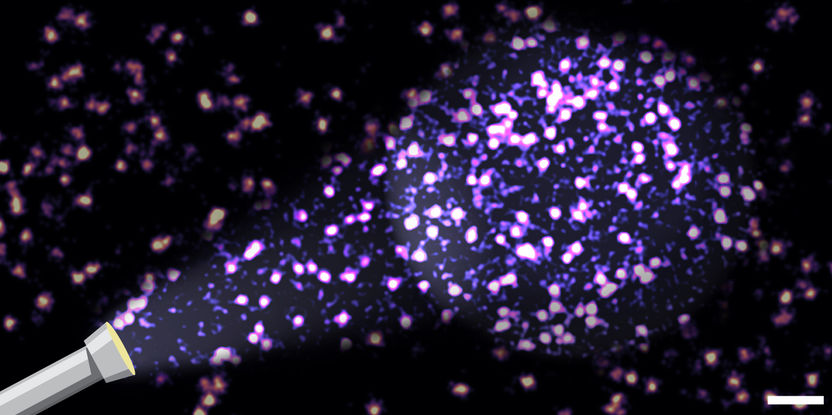
Localization clouds of individual adhesion proteins in cells. Many proteins remained undetectable when using conventional analytical methods. By using the new analytical method actual molecular parameters can be determined. Scale bar: 100 nm
© Lisa Fischer und Carsten Grashoff
Background and methodology
The attachment of cells is mediated by multi-molecular adhesion complexes that are built by hundreds of different proteins. The development of super-resolution microscopy, which was honoured with the Nobel Prize in 2014, allowed the identification of basic structural elements within such complexes. However, it remained unclear how individual proteins assemble and co-organize to form functional units. The laboratories of Prof. Dr. Carsten Grashoff at the Institute of Molecular Cell Biology (University of Münster) and Prof. Dr. Ralf Jungmann at the Max Planck Institute of Biochemistry (Munich) now developed a novel approach that allows the visualization and quantification of such molecular processes even in highly crowded subcellular structures.
“A substantial limitation even of the best super-resolution microscopy techniques is that many molecules remain undetected. It is therefore nearly impossible to make quantitative statements about processes of molecular complex formation in cells“, explains Lisa Fischer, PhD student in the Grashoff group and first author of the study. This difficulty could now be circumvented with a combination of experimental controls and theoretical considerations.
“By applying our new analytical method, we were able to provide evidence for the existence of a long suspected ternary adhesion complex. We knew already before that each of these three molecules is very important for cell adhesion”, explains Fischer. “However, it was not clear whether all three proteins come together to form a functional unit”. As the method is broadly applicable, the researchers believe that many other cellular processes will be studied with the new analysis procedure.
Original publication
Most read news
Original publication
L.S. Fischer, C. Klingner, T. Schlichthaerle, M.T. Strauss, R. Böttcher, R. Fässler, R. Jungmann, C. Grashoff; "Quantitative single-protein imaging reveals molecular complex formation of integrin, talin, and kindlin during cell adhesion"; Nature Communications; 12, 919 (2021).
Topics
Organizations
Other news from the department science

Get the life science industry in your inbox
By submitting this form you agree that LUMITOS AG will send you the newsletter(s) selected above by email. Your data will not be passed on to third parties. Your data will be stored and processed in accordance with our data protection regulations. LUMITOS may contact you by email for the purpose of advertising or market and opinion surveys. You can revoke your consent at any time without giving reasons to LUMITOS AG, Ernst-Augustin-Str. 2, 12489 Berlin, Germany or by e-mail at revoke@lumitos.com with effect for the future. In addition, each email contains a link to unsubscribe from the corresponding newsletter.


