Superresolution live-cell imaging provides unexpected insights into the dynamic structure of mitochondria
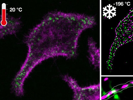
As power plants and energy stores, mitochondria are essential components of almost all cells in plants, fungi and animals. Until now, it has been assumed that these functions underlie a static structure of mitochondrial membranes. Researchers at the Heinrich Heine University Düsseldorf (HHU) and the University of California Los Angeles (UCLA) and have now discovered that the inner membranes of mitochondria are by no means static, but rather constantly changing their structure every few seconds in living cells. This dynamic adaptation process further increases the performance of our cellular power plants.
"In our opinion, this finding fundamentally changes the way our cellular power plants work and will probably change the textbooks," says Prof. Dr. Andreas Reichert, Institute of Biochemistry and Molecular Biology I at the HHU.
Mitochondria are extremely important components in cells performing vital functions including the regulated conversion of energy from food into chemical energy in the form of ATP. ATP is the energy currency of cells and an adult human being produces (and consumes) approximately 75 kilograms of ATP per day. One molecule of ATP is produced about 20,000 times a day and then consumed again for energy utilization. This immense synthesis capacity takes place in the inner membrane of the mitochondria, which has numerous folds called cristae. It was previously assumed that a specific static structure of the cristae ensured the synthesis of ATP. Whether and to what extent cristae membranes are able to dynamically adapt or alter their structure in living cells and which proteins are required to do so, was unknown.
The research team of Prof. Dr. Andreas Reichert with Dr. Arun Kondadi and Dr. Ruchika Anand from the Institute of Biochemistry and Molecular Biology I of the HHU in collaboration with the research team of Prof. Dr. Orian Shirihai and Prof. Dr. Marc Liesa from UCLA (USA) succeeded for the first time in showing that cristae membranes in living cells continuously change their structure dynamically within seconds within mitochondria. This showed that the cristae membrane dynamics requires a recently identified protein complex, the MICOS complex. Malfunctions of the MICOS complex can lead to various serious diseases, such as Parkinson's disease and a form of mitochondrial encephalopathy with liver damage. After the identification of the first protein component of this complex (Fcj1/Mic60) about ten years ago by Prof. Andreas Reichert and his research group, this is another important step to elucidate the function of the MICOS complex.
"Our now published observations lead to the model that cristae, after membrane fission, can exist for a short time as isolated vesicles within mitochondria and then re-fuse with the inner membrane. This enables an optimal and extremely rapid adaptation to the energetic requirements in a cell," said Prof. Andreas Reichert.
Other news from the department science
Most read news
More news from our other portals
See the theme worlds for related content
Topic World Cell Analysis
Cell analyse advanced method allows us to explore and understand cells in their many facets. From single cell analysis to flow cytometry and imaging technology, cell analysis provides us with valuable insights into the structure, function and interaction of cells. Whether in medicine, biological research or pharmacology, cell analysis is revolutionizing our understanding of disease, development and treatment options.


