Tracking the HI virus
Researchers makes visible, how AIDS pathogens multiply in the body
An international team of researchers led by Dr Cyril Favard and Dr Delphine Muriaux from the Montpellier Infectious Disease Research Institute in collaboration with Prof. Dr Christian Eggeling from the Friedrich Schiller University Jena, the Leibniz Institute of Photonic Technology (Leibniz IPHT), and the University of Oxford has succeeded in using high-resolution imaging to make visible how the HI virus spreads between living cells and which molecules it requires for this. Using superresolution STED fluorescence microscopy, the researchers provide direct proof for the first time that the AIDS pathogen creates a certain lipid environment for replication. "We have thus created a method for investigating how this multiplication can potentially be prevented," says Christian Eggeling.

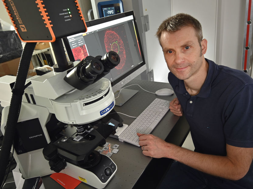
Christian Eggeling from Leibniz IPHT and the Friedrich Schiller University Jena
Jan-Peter Kasper/ Friedrich-Schiller-Universität Jena
The researchers focused on the sluice through which the HI virus (Human Immunodeficiency Virus) emerges from the cell after having infected it: the plasma membrane of the host cell. They used the protein Gag as a marker, which coordinates the processes involved in the maturation of the virus. "Where this protein accumulates, the decisive processes take place that lead to the virus releasing itself and infecting other cells," explains Christian Eggeling. In order to decipher these, the researchers examined the diffusion at this budding site of the virus particle. They found out that only certain lipids interact with the HI virus. Although these lipids were already known in principle, the research team was able to prove this interaction directly in living and infected cells for the first time.
Point of attack to prevent the virus from multiplying
"This provides us with a potential target for antiviral drugs," says Christian Eggeling. "Knowing which molecules the HI virus needs in order to leave the cell and multiply is a crucial prerequisite for investigating how this can be prevented. With our technology, we can now follow this directly.“ Christian Eggeling and his team now want to develop antibodies that attack precisely these molecules — and thus suppress the spread of the virus.
"We not only want to study these antibodies from a medical point of view, but also to find out how their biophysical interaction can be used to enhance their efficacy," says Eggeling, describing his research program. "For this purpose, we analyze biological processes — namely the interaction of cells and molecules - with the aid of physical parameters such as diffusion. A good year ago, the physicist moved from Oxford to Jena. In addition to his professorship for "Superresolution Microscopy" at the university, he heads the research department "Biophysical Imaging" at Leibniz IPHT. He also leads his research group at the MRC Human Immunology Unit and at the Wolfson Imaging Centre of the Weatherall Institute of Molecular Medicine at Oxford University.
Christian Eggeling combines spatially superresolution fluorescence microscopy techniques with methods that enable the movement of labelled molecules to be tracked in real time in order to understand how diseases develop at the smallest molecular level. This enables him and his team of researchers to investigate individual molecules - for example in cell membranes - in living cells spatially and temporally. "This enables us to reveal cellular mechanisms at the molecular level that are far too fast for previous investigation methods and run on spatial scales that are far too small.
Christian Eggeling has already researched new superresolution fluorescence microscopy techniques at the Max Planck Institute for Biophysical Chemistry in Göttingen in the group of Stefan W. Hell. Together with Eric Betzig and William E. Moerner, Stefan Hell received the Nobel Prize for Chemistry in 2014. In Jena, Eggeling is now working closely with biologists and physicians to find out how these methods can be used to detect diseases earlier and more accurately and possibly even prevent them.
Original publication
Other news from the department science
Most read news
More news from our other portals
See the theme worlds for related content
Topic world Fluorescence microscopy
Fluorescence microscopy has revolutionized life sciences, biotechnology and pharmaceuticals. With its ability to visualize specific molecules and structures in cells and tissues through fluorescent markers, it offers unique insights at the molecular and cellular level. With its high sensitivity and resolution, fluorescence microscopy facilitates the understanding of complex biological processes and drives innovation in therapy and diagnostics.


