Researchers combine light and X-ray microscopy for comprehensive insights
Researchers at the University of Goettingen have used a novel microscopy method. In doing so they were able to show both the illuminated and the "dark side" of the cell.

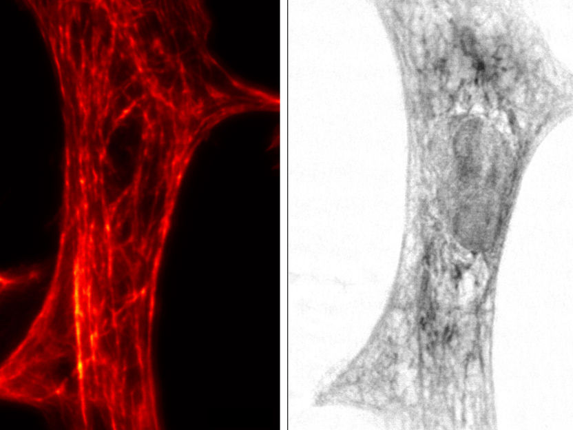
STED image (left) and x-ray imaging (right) of the same cardiac tissue cell from a rat.
University of Goettingen
The team led by Prof. Dr. Tim Salditt and Prof. Dr. Sarah Köster from the Institute of X-Ray Physics "attached" small fluorescent markers to the molecules of interest, for example proteins or DNA. The controlled switching of the fluorescent dye in the so-called STED (Stimulated Emission Depletion) microscope then enables highest resolution down to a few billionth of a meter. The Goettingen team combined a light microscope according to the STED principle, which represents the illuminated area of the cell, with an X-ray microscope, which represents the unilluminated area of the cell.
"We used the innovative microscope to show a network of protein filaments in heart muscle cells," explains Marten Bernhardt, first author of the publication. "The protein networks contained therein were mapped in STED mode. We were then able to fit these STED images into the X-ray images of the cell. Both recordings are recorded practically directly one after another," says Bernhardt. "We hope that the complementary contrasts will provide us with a more complete understanding of the contraction of heart muscle cells and their generation of strength," adds Salditt. In designing the STED microscope, the scientists worked closely with the research centre “Deutsches Elektronen-Synchrotron DESY” and the Abberior company founded by Goettingen Nobel Prize winner Prof. Dr. Stefan W. Hell. "In future, we also want to observe dynamic processes in living cells," concludes Köster.
Original publication
Other news from the department science
Most read news
More news from our other portals
See the theme worlds for related content
Topic world Fluorescence microscopy
Fluorescence microscopy has revolutionized life sciences, biotechnology and pharmaceuticals. With its ability to visualize specific molecules and structures in cells and tissues through fluorescent markers, it offers unique insights at the molecular and cellular level. With its high sensitivity and resolution, fluorescence microscopy facilitates the understanding of complex biological processes and drives innovation in therapy and diagnostics.


