Scientists gain new understanding of disease-causing bacteria
For the first time, research elucidates the cellular structure of syphilis pathogen
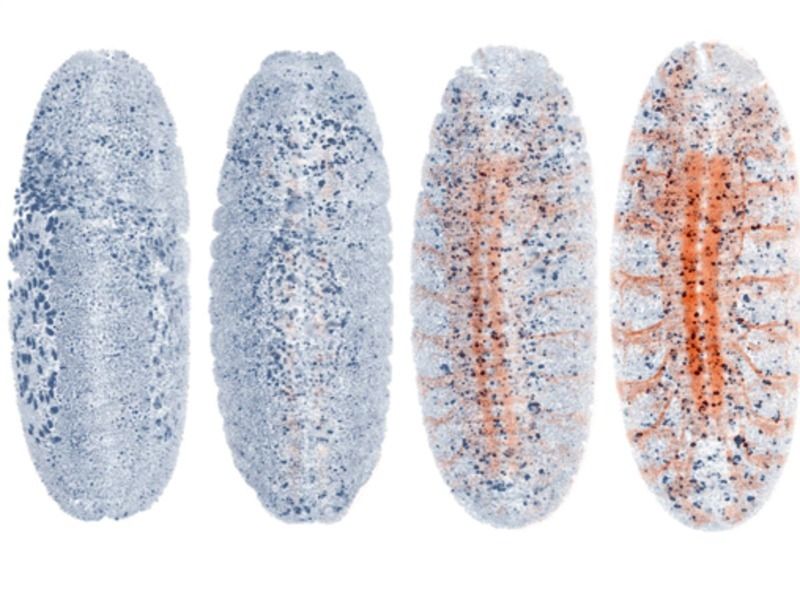
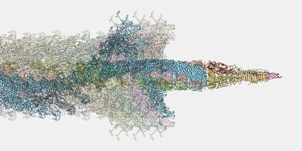
A team of scientists from The Forsyth Institute, the University of Connecticut Health Center, the CDC and the Wadsworth Center, have used state-of-the-art technology to elucidate the molecular architecture of Treponema pallidum, the bacterium which causes syphilis. The previously unknown detailed structure of the bacteria can now be shown in three dimensions. This provides the first real image of the pathogen and reveals previously unknown features, which may help fight the spread of syphilis. cryo-electron tomography (CET) is a type of microscope that is used to obtain a three-dimensional reconstruction of a sample from two dimensional images at extremely low temperatures. Using CET, the research team has clarified the fundamental differences between Treponema pallidum and other gram-negative bacteria. This research will be featured in the Journal of Bacteriology. According to lead author Jacques Izard, Ph.D., this work provides a clear snapshot of a cell in real time. Added Izard, "This changes how we study this bacterium. Having an accurate architecture of the cell provides important insight for understanding how it becomes invasive in the human body. With this information we may learn how to stop disease progression." Over a decade ago, the publication of the Treponema pallidum genome sequence provided a much needed parts list for the bacterium. However, scientists have learned very little about how these components are organized to create this extremely virulent and immuno-evasive pathogen. CET has emerged as a powerful tool for bridging the knowledge gap. With this technique, thin films of cells are frozen to preserve cell structure in a close-to-native state, avoiding degradation caused by preparation for traditional microscopy. A series of images acquired as the sample is progressively tilted in an electron microscope are used to generate a 3D image. With CET T. pallidum cells appeared to form flat waves and did not contain an outer coat. This highly motile organism can attach to human cells by its tip. The present work has shown that the tip of this bacteria has a unique structure among pathogens, which improved the understanding of cell attachment and tissue penetration. Additionally, novel structural evidence explains how those bacteria mysteriously move with the flagella inside their cell body.
Other news from the department science


Most read news


More news from our other portals


Last viewed contents
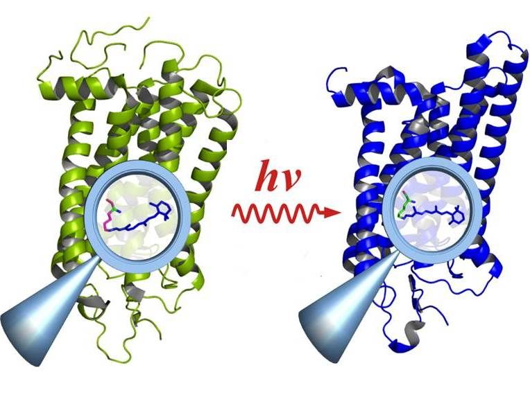
Watching complex molecules at work
Solipsism_syndrome
Pinolenic_acid
Allopregnanolone
Joseph_von_Gerlach
Cross species transfer of genes has driven evolution
Histology