Decoding cell division's mysterious spindle matrix
It's known that the act of chromosome separation is facilitated by a structure called the spindle, a framework composed of interconnected fibrous microtubules. The two copies of each chromosome (made through DNA replication) are attached to the spindle's microtubule fibers and then moved apart to opposite sides of the dividing cell. In this way each of the two daughter cells receives one complete copy of the genetic material. This process requires the microtubule fibers to be organized into a spindle-like structure. Besides the microtubule fibers, the spindle also contains other densely packed materials that are visible under the electron microscope. These poorly understood materials are referred to as the spindle matrix and are believed to be important for cell division.
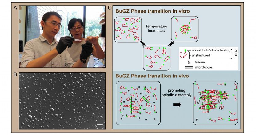
Concentrated cell lysates can be made by spinning the mature Xenopus eggs in a centrifuge without buffer, which crushes and stratifies the cell contents. The stratified golden layer above the needle in panel A is an undiluted cell lysate that supports many reactions found in cells including the assembly of the spindle apparatus in a test tube. This system has allowed the investigators to discover a spindle matrix protein called BuGZ that uses its self-association droplet-forming property shown in panel B to support the assembly of spindle and its matrix as depicted in panel C.
Yixian Zheng and Hao Jiang
New work from a team led by Carnegie's Yixian Zheng zeroes in on one particular spindle matrix protein, called BuGZ. The team's findings show that BuGZ supports the assembly of the spindle matrix and microtubules.
The team found that under spindle-assembly conditions, many copies of the BuGZ protein condense along the microtubule fibers to form droplets. This condensation occurs because molecules of the BuGZ protein can assemble with each other, and is referred to as phase transition. During mitosis, these condensed BuGZ droplets promote the assembly of the spindle matrix, which makes additional microtubule fibers and links the fibers together, thereby promoting assembly of the spindle itself.
The formation of BuGZ droplets is the result of an evolutionarily conserved water-repelling (or hydrophobic) segment of BuGZ, which rejects interaction with the surrounding cellular fluid. Instead BuGZ proteins self-associate into these "inward-looking" droplet shapes. The team found that several other proteins found in the spindle matrix in both frogs and fruit flies have segments that resemble the water-repelling portion of BuGZ. They speculate that the whole spindle matrix could be formed through self-association of the hydrophobic segments of these proteins. This could help explain why many seemingly unrelated proteins make up the spindle matrix, which has been one of the central mysteries of the cell-division process.
"Because BuGZ is evolutionarily conserved and found in both vertebrates and invertebrates, our findings should open the door to further research on spindles and their matrices in a wide variety of organisms," Zheng said.
Most read news
Organizations
Other news from the department science

Get the life science industry in your inbox
By submitting this form you agree that LUMITOS AG will send you the newsletter(s) selected above by email. Your data will not be passed on to third parties. Your data will be stored and processed in accordance with our data protection regulations. LUMITOS may contact you by email for the purpose of advertising or market and opinion surveys. You can revoke your consent at any time without giving reasons to LUMITOS AG, Ernst-Augustin-Str. 2, 12489 Berlin, Germany or by e-mail at revoke@lumitos.com with effect for the future. In addition, each email contains a link to unsubscribe from the corresponding newsletter.
